Copyright
©The Author(s) 2025.
World J Psychiatry. Jun 19, 2025; 15(6): 105751
Published online Jun 19, 2025. doi: 10.5498/wjp.v15.i6.105751
Published online Jun 19, 2025. doi: 10.5498/wjp.v15.i6.105751
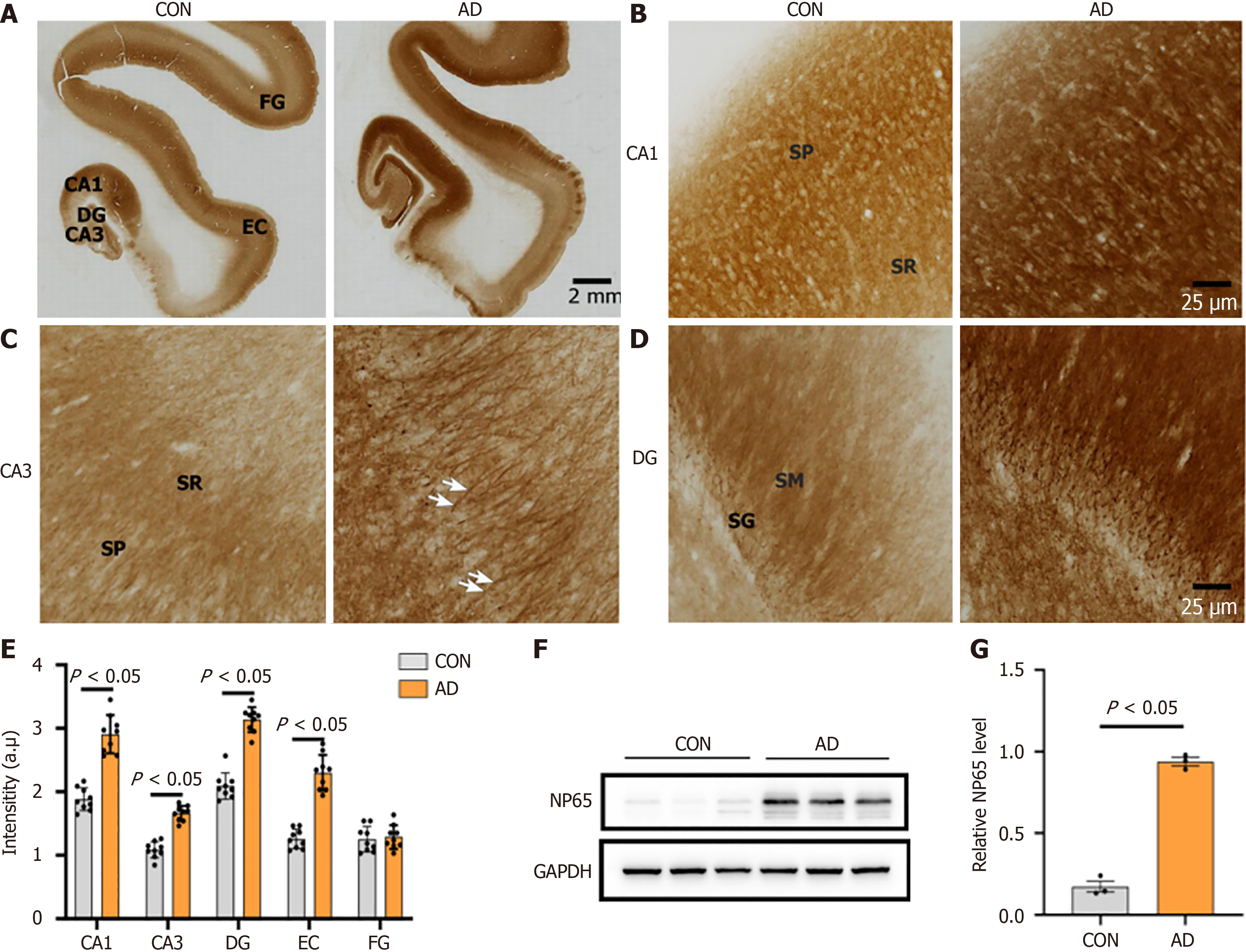
Figure 5 Neuroplastin 65 immunoreactivity pattern in the hippocampal formation and temporal cortex derived from individuals with Alzheimer’s disease and age-matched controls.
A: Low-power microphotographs display the regional distribution of neuroplastin 65 (NP65) in the hippocampal formation, entorhinal cortex and fusiform gyrus of controls (CON) and Alzheimer’s disease (AD) (scale bars = 2 mm); B-D: High-power microphotographs of cornu ammonis (CA) 1, CA3 and dentate gyrus, respectively. Note the strong positive staining of apical dendrites of the stratum pyramidale of CA3 (indicated by arrow in C) in AD (scale bars = 50 μm); E: Quantification of NP65 immunoreactivity in the hippocampal formation, entorhinal cortex and fusiform gyrus between AD and CON; F and G: NP65 immunoreactive bands from total membrane protein of the hippocampus in AD and CON, and quantitative analysis showing NP65 levels in the hippocampus of AD and CON (data from two independent experiments). NP65: Neuroplastin 65; DG: Dentate gyrus; CA: Cornu ammonis; EC: Entorhinal cortex; SG: Stratum granulosum; SM: Stratum moleculare; SR: Stratum radiatum; SP: Stratum pyramidale; FG: Fusiform gyrus; GAPDH: Glyceraldehyde-3-phosphate dehydrogenase; Neuroplastin 65 (NP65); CON: Controls; AD: Alzheimer’s disease.
- Citation: Zheng YN, Wang Y, Chen L, Xu LZ, Zhang L, Wang JL, Liu J, Zhang QL, Yuan QL. Increased expression of the neuroplastin 65 protein is involved in neurofibrillary tangles and amyloid beta plaques in Alzheimer’s disease. World J Psychiatry 2025; 15(6): 105751
- URL: https://www.wjgnet.com/2220-3206/full/v15/i6/105751.htm
- DOI: https://dx.doi.org/10.5498/wjp.v15.i6.105751


