Copyright
©The Author(s) 2025.
World J Psychiatry. Jun 19, 2025; 15(6): 105751
Published online Jun 19, 2025. doi: 10.5498/wjp.v15.i6.105751
Published online Jun 19, 2025. doi: 10.5498/wjp.v15.i6.105751
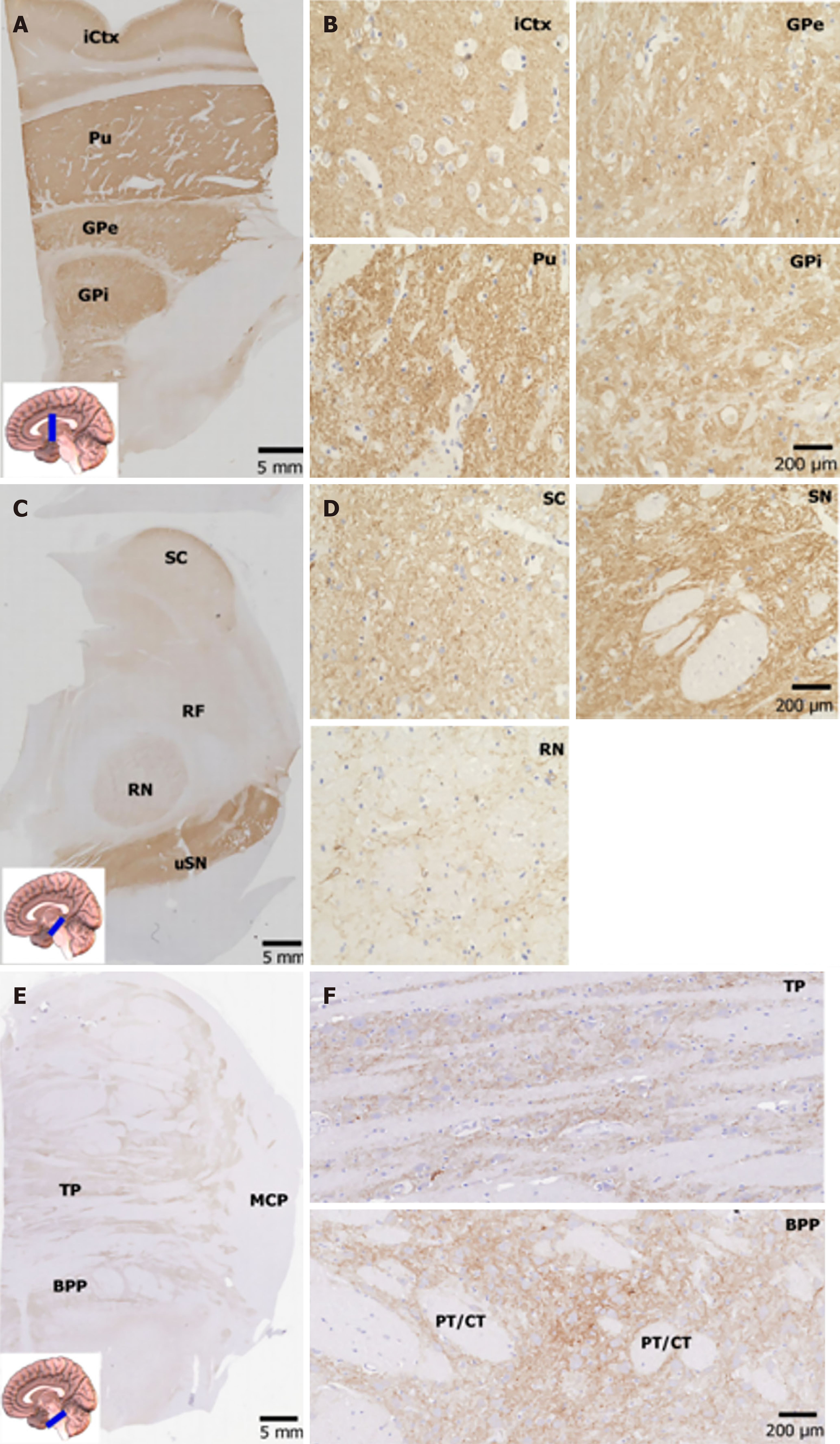
Figure 3 Expression of neuroplastin 65 immunoreactivity in human lentiform nucleus, midbrain and pons.
A: A representative immunohistochemical staining micrograph in the lentiform nucleus, with an inset indicating the position of the sampling (scale bars = 5 mm); B: A representative immunohistochemical staining micrograph in the lentiform nucleus, with an inset indicating the position of the sampling (higher power views, scale bars = 200 μm). Note that neuroplastin 65 immunoreactivity shows strong punctate staining in the putamen, while there are dense networks in the globus pallidus; C: An immunohistochemical staining micrograph in the midbrain at a low magnification view, with an inset indicating the position of the sampling (scale bars = 5 mm); D: An immunohistochemical staining micrograph in the midbrain at a low magnification view, with an inset indicating the position of the sampling (higher power view, scale bars = 200 μm). Note that there is positive punctate staining and fibers in the substantia nigra and less intense positive fibers in the red nucleus; E: A low magnification view of the pons, with an inset indicating the position of the sampling (scale bars = 5 mm); F: A low magnification view of the pons, with an inset indicating the position of the sampling (higher power views, scale bars = 200 μm). The nucleus is stained by hematoxylin. iCtx: Insular cortex; Pu: Putamen; GPe: External part of the globus pallidus; GPi: Internal part of the globus pallidus; IC: Internal capsule; CC: Crus cerebri; SN: Substantia nigra; RN: Red nucleus; SC: Superior colliculus; RF: Reticular formation; BPP: Basilar part of the pons; TP: Tegmentum of the pons; PT: Pyramidal tract; CT: Corticopontine tract; MCP: Middle cerebellar peduncle.
- Citation: Zheng YN, Wang Y, Chen L, Xu LZ, Zhang L, Wang JL, Liu J, Zhang QL, Yuan QL. Increased expression of the neuroplastin 65 protein is involved in neurofibrillary tangles and amyloid beta plaques in Alzheimer’s disease. World J Psychiatry 2025; 15(6): 105751
- URL: https://www.wjgnet.com/2220-3206/full/v15/i6/105751.htm
- DOI: https://dx.doi.org/10.5498/wjp.v15.i6.105751


