Copyright
©The Author(s) 2025.
World J Psychiatry. Jun 19, 2025; 15(6): 105751
Published online Jun 19, 2025. doi: 10.5498/wjp.v15.i6.105751
Published online Jun 19, 2025. doi: 10.5498/wjp.v15.i6.105751
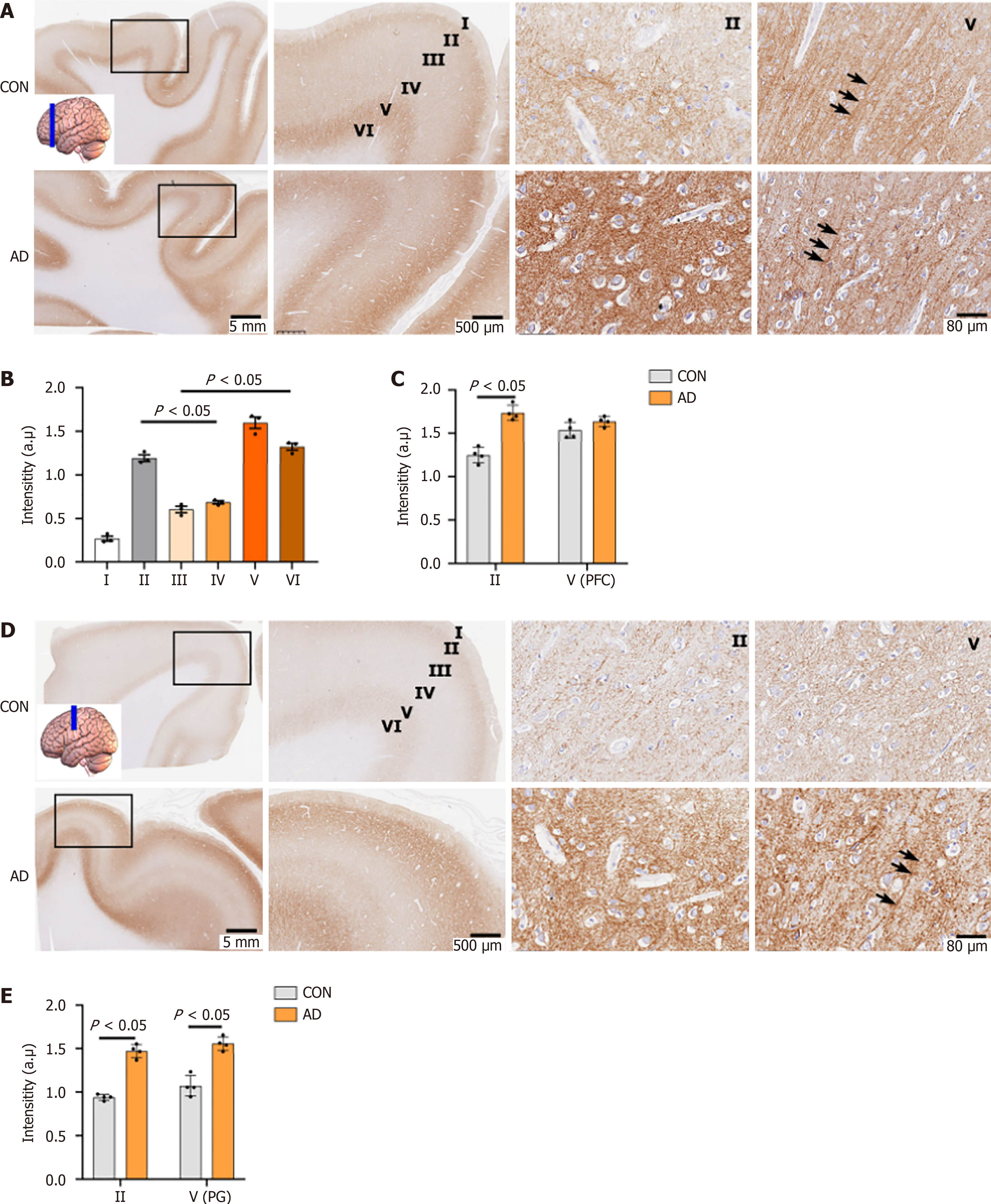
Figure 2 Neuroplastin 65 immunoreactive pattern in the dorsolateral frontal cortex of individuals with Alzheimer’s disease and age-matched controls.
A: Representative Neuroplastin 65 (NP65) immunohistochemical staining micrographs of a coronal section in the dorsolateral prefrontal cortex (dPFC) from controls (CON) and Alzheimer’s disease (AD), with an inset indicating the sampling position. Note the strongly positive apical dendrites (indicated by an arrow) in layer V; B: Quantification of NP65 immunoreactivity in sublayers of the dPFC in CON; C: Comparison of NP65 immunoreactivity in layer II and V of the dPFC between AD and CON; D: Representative NP65 immunohistochemical staining micrographs of a coronal section in the precentral gyrus of CON and AD, with an inset indicating the sampling position. Note the strongly positive apical dendrites (indicated by an arrow) in layer V of the AD brain; E: Comparison of NP65 immunoreactivity in layer II and V of the precentral gyrus between AD and CON. The nucleus is stained with hematoxylin. PFC: Prefrontal cortex; CON: Controls; AD: Alzheimer’s disease; PG: Precentral gyrus.
- Citation: Zheng YN, Wang Y, Chen L, Xu LZ, Zhang L, Wang JL, Liu J, Zhang QL, Yuan QL. Increased expression of the neuroplastin 65 protein is involved in neurofibrillary tangles and amyloid beta plaques in Alzheimer’s disease. World J Psychiatry 2025; 15(6): 105751
- URL: https://www.wjgnet.com/2220-3206/full/v15/i6/105751.htm
- DOI: https://dx.doi.org/10.5498/wjp.v15.i6.105751


