Copyright
©The Author(s) 2021.
World J Psychiatr. Oct 19, 2021; 11(10): 681-695
Published online Oct 19, 2021. doi: 10.5498/wjp.v11.i10.681
Published online Oct 19, 2021. doi: 10.5498/wjp.v11.i10.681
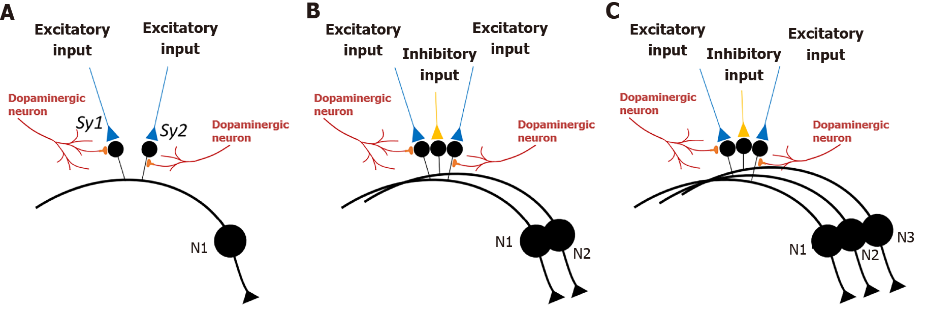
Figure 3 Interactions between spines of medium spiny neurons that synapse with excitatory inputs and spines of medium spiny neurons that synapse with inhibitory inputs.
A: Adjacent spines (small black circles) on the dendrite of a medium spiny neuron (MSN) (N1) (cell body is drawn in a large black circle) that synapse with two excitatory inputs (in blue) to form synapses Sy1 and Sy2). Golgi staining shows that spines are physically well separated from each other on the dendrites of MSNs[23,24] such that the inter-spine space is occupied by spines of other dendrites or processes of other neurons or glial cells. This increases the probability that the nearest spine to a spine on the dendrite of a MSN is most likely a spine that belongs to another neuron, or in rare cases belongs to another branch of the same neuron. Note that dopaminergic inputs synapse either onto the head or neck region of spines that synapse with excitatory inputs; B: In between two adjacent spines of MSN N1 shown in figure A, there is a spine that belongs to a second MSN (N2). This spine synapses with an inhibitory input (in orange). All the spines are electrically insulated from each other by fluid extracellular matrix. Natural stimulants or cocaine abuse causes release of dopamine that will cause enlargement of spines that synapse with excitatory inputs. Since the spine that synapses with the inhibitory input is spatially interposed between the expanding spines, inter-postsynaptic functional LINKs are formed between those three spines; C: Same configuration of two spines of MSNs that synapse with excitatory inputs and one middle spine synapsing with inhibitory input. Here, these spines belong to three different MSNs.
- Citation: Vadakkan KI. Framework for internal sensation of pleasure using constraints from disparate findings in nucleus accumbens. World J Psychiatr 2021; 11(10): 681-695
- URL: https://www.wjgnet.com/2220-3206/full/v11/i10/681.htm
- DOI: https://dx.doi.org/10.5498/wjp.v11.i10.681


