Copyright
©2013 Baishideng Publishing Group Co.
World J Med Genet. Nov 27, 2013; 3(4): 41-54
Published online Nov 27, 2013. doi: 10.5496/wjmg.v3.i4.41
Published online Nov 27, 2013. doi: 10.5496/wjmg.v3.i4.41
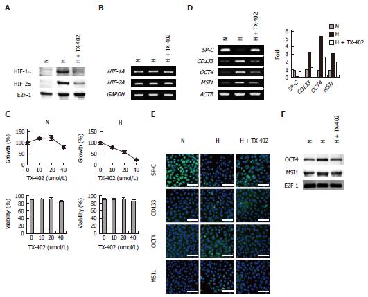
Figure 8 Effects of TX-402 on hypoxia-inducible factor-α expression, cell proliferation, viability and gene expression levels of A549 cells.
The cells were cultured under normoxic (N) or hypoxic (H) conditions in the presence or absence of 20 μmol/L TX-402 for 3 d; A: Expression of HIF-α subunits, Nuclear extracts and total RNA were subjected to Western blot; B: Expression of HIF-α subunits, Nuclear extracts and total RNA were subjected to reverse transcription-polymerase chain reaction (RT-PCR) analysis; C: Cell growth and viability, bars, SD (n = 3); D: RT-PCR analysis of the expression of SP-C, CD133, OCT4 and MSI1 Mrna, the expression level of each gene was normalized to that of β-Actin (ACTB), data are shown as fold-change relative to normoxia (normoxia values set to equal 1); E: Immunofluorescence analysis of the expression of SP-C, CD133, OCT4 and MSI1 protein, Nuclei were stained with DAPI, and the merged images are shown, Perinuclear and nuclear localization of OCT4 and MSI1 was observed, Scale bars = 50 μm; F: Western blot analysis of the nuclear localization of OCT4 and MSI1, Nuclear extracts of the cells were subjected to the analysis, E2F-1 was used as a loading control.
- Citation: Akimoto M, Nagasawa H, Hori H, Uto Y, Honma Y, Takenaga K. An inhibitor of HIF-α subunit expression suppresses hypoxia-induced dedifferentiation of human NSCLC into cancer stem cell-like cells. World J Med Genet 2013; 3(4): 41-54
- URL: https://www.wjgnet.com/2220-3184/full/v3/i4/41.htm
- DOI: https://dx.doi.org/10.5496/wjmg.v3.i4.41


