Copyright
©The Author(s) 2022.
World J Clin Infect Dis. Sep 29, 2022; 12(2): 69-75
Published online Sep 29, 2022. doi: 10.5495/wjcid.v12.i2.69
Published online Sep 29, 2022. doi: 10.5495/wjcid.v12.i2.69
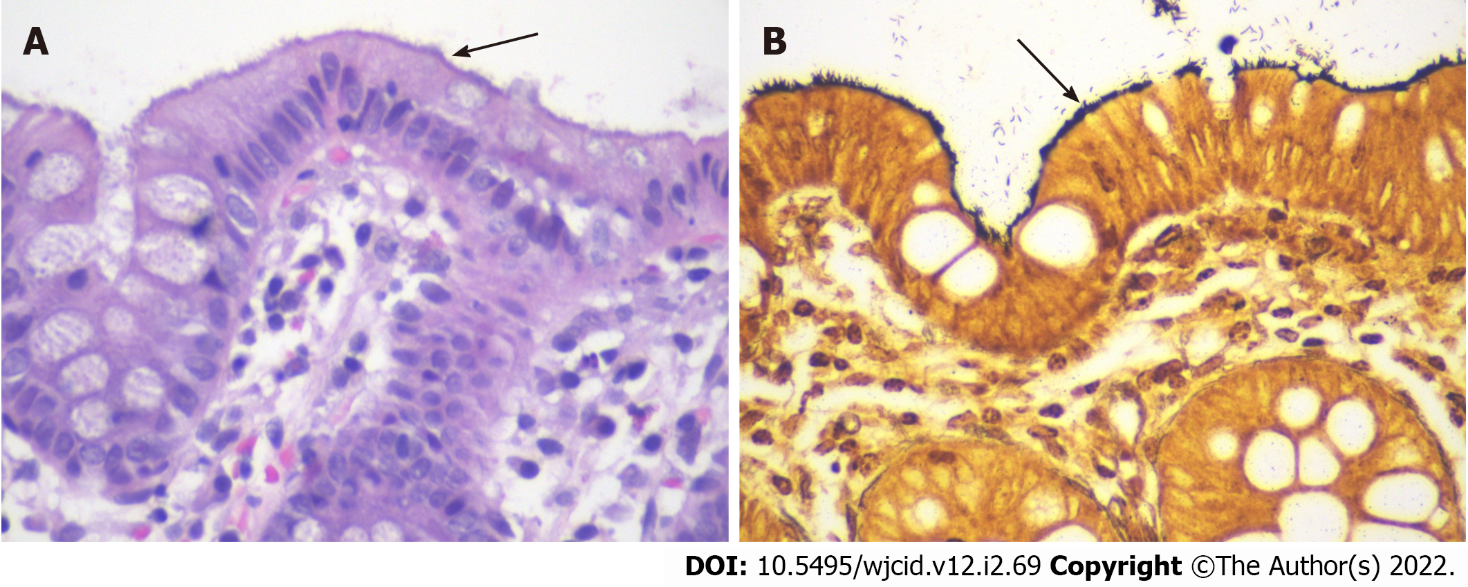
Figure 2 Pathology images of samples taken during colonoscopy.
A: Hematoxylin and eosin stain (× 600 magnification) showed intestinal spirochetosis appearing as a basophilic, fuzzy lining over the luminal surface of colonocytes (“false brush border”); B: Steiner stain (× 600 magnification) highlighted spirochetes overlying the luminal surface.
- Citation: Novotny S, Mizrahi J, Yee EU, Clores MJ. Incidental diagnosis of intestinal spirochetosis in a patient with chronic hepatitis B: A case report. World J Clin Infect Dis 2022; 12(2): 69-75
- URL: https://www.wjgnet.com/2220-3176/full/v12/i2/69.htm
- DOI: https://dx.doi.org/10.5495/wjcid.v12.i2.69


