Copyright
©The Author(s) 2015.
World J Hypertens. May 23, 2015; 5(2): 41-52
Published online May 23, 2015. doi: 10.5494/wjh.v5.i2.41
Published online May 23, 2015. doi: 10.5494/wjh.v5.i2.41
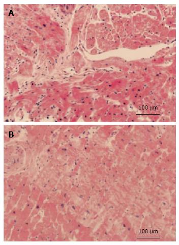
Figure 6 Representative cross sections of myocardial biopsy specimens.
A: Hypertrophic cardiomyopathy showing disorganized arrangement of hypertrophic myocytes; B: Hypertensive cardiomyopathy patients showing parallel alignment of hypertrophic myocytes. Sections were stained with hematoxylin-eosin. (Reproduced from Kato et al[62], 2004).
- Citation: Kuroda K, Kato TS, Amano A. Hypertensive cardiomyopathy: A clinical approach and literature review. World J Hypertens 2015; 5(2): 41-52
- URL: https://www.wjgnet.com/2220-3168/full/v5/i2/41.htm
- DOI: https://dx.doi.org/10.5494/wjh.v5.i2.41


