
















Copyright: ©Author(s) 2026.
World J Exp Med. Mar 20, 2026; 16(1): 112270
Published online Mar 20, 2026. doi: 10.5493/wjem.v16.i1.112270
Published online Mar 20, 2026. doi: 10.5493/wjem.v16.i1.112270
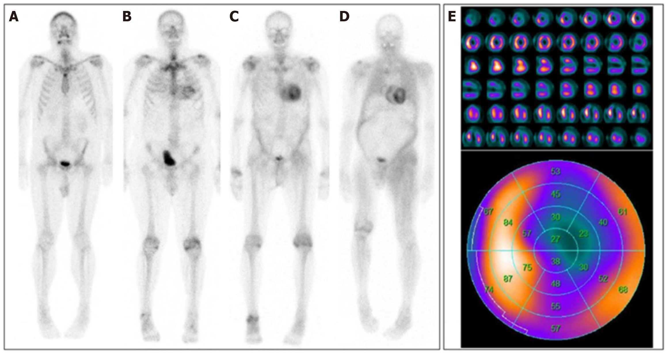
Figure 3 Planar 99mTc-DPD scintigraphies of four different patients.
A: No cardiac uptake (Perugini score 0); B: Light cardiac uptake with preserved delineation of bone tissue (Perugini score 1); C: Strong cardiac uptake above that of bone tissue and increased soft-tissue uptake, particularly in the shoulder, abdominal wall, and gluteal region (Perugini score 2); D: Strong cardiac and soft-tissue uptake with no discernable bone-tissue uptake, suggesting difuse amyloid soft-tissue infltration (Perugini score 3); E: Single-photon emission computed tomography imaging: Short axis, vertical long axis, and horizontal long axis slices at the top and the corresponding polar plot at the bottom; white/yellow indicates high uptake; blue/black indicates little or no uptake[38]. Citation: Yilmaz A, Bauersachs J, Bengel F, Büchel R, Kindermann I, Klingel K, Knebel F, Meder B, Morbach C, Nagel E, Schulze-Bahr E, Aus dem Siepen F, Frey N. Diagnosis and treatment of cardiac amyloidosis: Position statement of the German Cardiac Society (DGK). Clin Res Cardiol 2021; 110: 479-506. Copyright© The Authors 2021. Published by Springer Nature. The article is open access (Supplementary material).
- Citation: Bouziane M, Moufid O, Habbal R. Cardiac amyloidosis: From diagnosis to therapeutics breakthroughs. World J Exp Med 2026; 16(1): 112270
- URL: https://www.wjgnet.com/2220-315x/full/v16/i1/112270.htm
- DOI: https://dx.doi.org/10.5493/wjem.v16.i1.112270