
















Copyright: ©Author(s) 2026.
World J Exp Med. Mar 20, 2026; 16(1): 112270
Published online Mar 20, 2026. doi: 10.5493/wjem.v16.i1.112270
Published online Mar 20, 2026. doi: 10.5493/wjem.v16.i1.112270
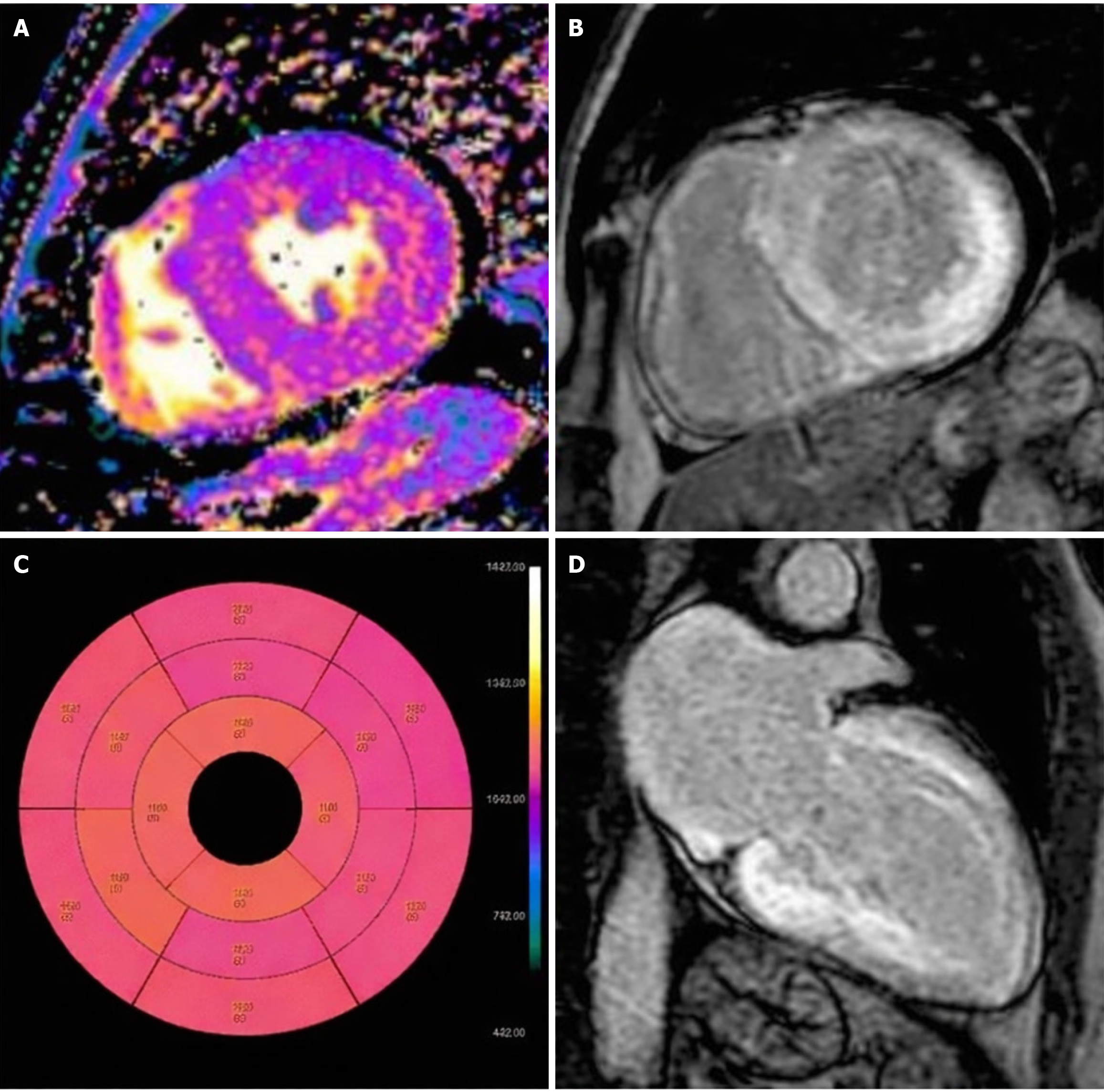
Figure 2 Patient diagnosed with endomyocardial biopsy.
A: T1 native mapping demonstrates abnormally high values in all entire left ventricle (LV) walls in cardiac amyloidosis; B: Diffuse transmural LV and subendocardial right ventricle (RV) late gadolinium enhancement (LGE) in two-chamber short; C: Bulls-eye map of T1 native; D: Diffuse transmural LV and subendocardial RV LGE in long axes[26]. Citation: Maggialetti N, Torrente A, Lorusso G, Villanova I, Ficco M, Gravina M, Ferrari C, Giordano L, Granata V, Rubini D, Lucarelli NM, Stabile Ianora AA, Scardapane A. Role of Cardiovascular Magnetic Resonance in Cardiac Amyloidosis: A Narrative Review. J Pers Med 2024; 14: 407. Copyright© The Authors 2024. Published by MDPI. The article is open access (Supplementary material).
- Citation: Bouziane M, Moufid O, Habbal R. Cardiac amyloidosis: From diagnosis to therapeutics breakthroughs. World J Exp Med 2026; 16(1): 112270
- URL: https://www.wjgnet.com/2220-315x/full/v16/i1/112270.htm
- DOI: https://dx.doi.org/10.5493/wjem.v16.i1.112270