Copyright
©The Author(s) 2021.
World J Exp Med. May 20, 2021; 11(3): 30-36
Published online May 20, 2021. doi: 10.5493/wjem.v11.i3.30
Published online May 20, 2021. doi: 10.5493/wjem.v11.i3.30
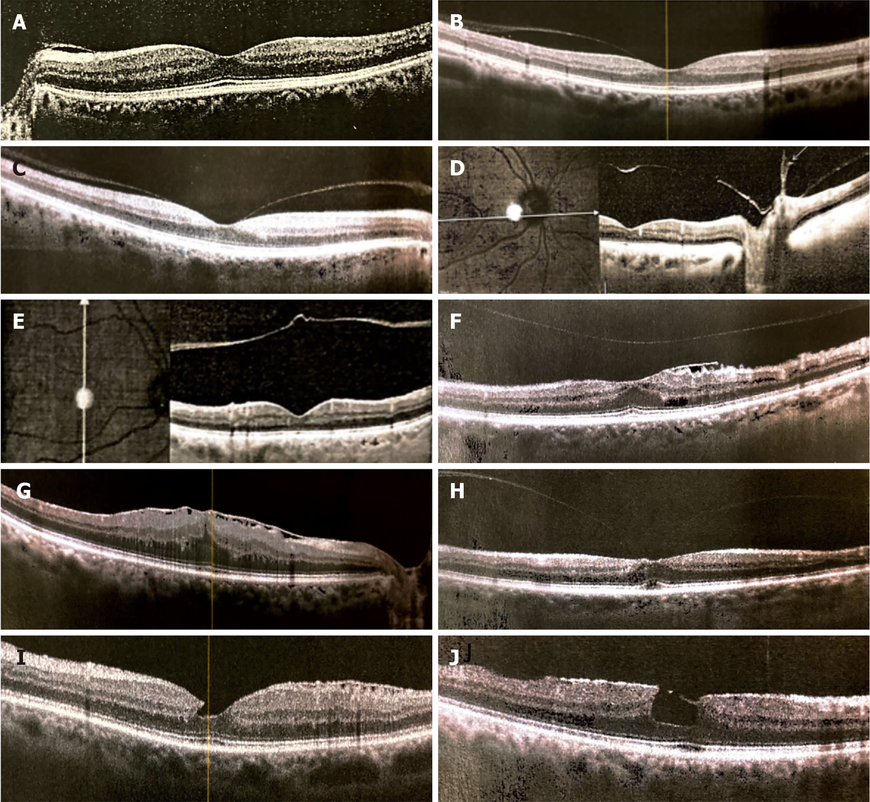
Figure 2 Different phases of posterior vitreous detachment visualized by optical coherence tomography macula scans.
A-D: Various stages of posterior vitreous detachment (PVD) can be seen in focal perifoveal sectors (A-C) and around the optic nerve head (D); E: Complete PVD is seen when a crumpled translucent membrane appears floating in mid vitreous; F-J: Results of abnormal PVD caused by vitreoretinal adherence and traction can lead to macular pucker (F and G) and macular lamellar holes (H-J).
- Citation: Ramovecchi P, Salati C, Zeppieri M. Spontaneous posterior vitreous detachment: A glance at the current literature. World J Exp Med 2021; 11(3): 30-36
- URL: https://www.wjgnet.com/2220-315x/full/v11/i3/30.htm
- DOI: https://dx.doi.org/10.5493/wjem.v11.i3.30


