Copyright
©The Author(s) 2019.
World J Exp Med. Dec 19, 2019; 9(2): 14-31
Published online Dec 19, 2019. doi: 10.5493/wjem.v9.i2.14
Published online Dec 19, 2019. doi: 10.5493/wjem.v9.i2.14
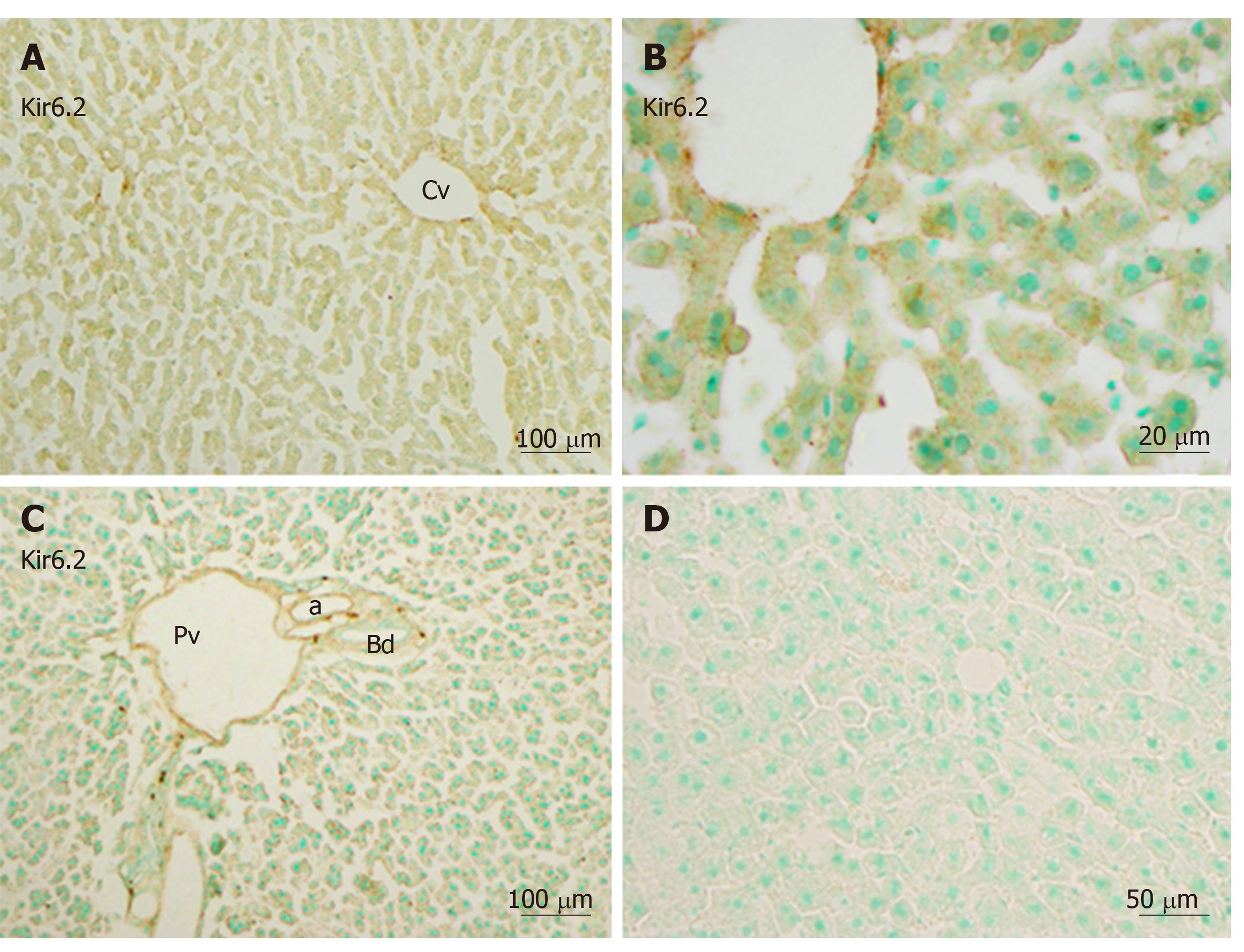
Figure 3 Immunoreactivity with Kir6.
2 was expressed in hepatocytes and sinusoidal cells of the liver. A and B: Mediated immunoreactivity was seen in the area around the central vein; A and C: Mediated immunoreactivity was weaker in the border areas; D: A negative control section devoid of staining (no more than background staining was observed). a: Artery; Bd: Bile duct, Cv: Central vein; Pv: Portal vein. Bars: 100 µm (A and C), 20 µm (B), 50 µm (D).
- Citation: Zhou M, Yoshikawa K, Akashi H, Miura M, Suzuki R, Li TS, Abe H, Bando Y. Localization of ATP-sensitive K+ channel subunits in rat liver. World J Exp Med 2019; 9(2): 14-31
- URL: https://www.wjgnet.com/2220-315X/full/v9/i2/14.htm
- DOI: https://dx.doi.org/10.5493/wjem.v9.i2.14


