
















Copyright: ©Author(s) 2026.
World J Crit Care Med. Sep 9, 2026; 15(3): 117716
Published online Sep 9, 2026. doi: 10.5492/wjccm.117716
Published online Sep 9, 2026. doi: 10.5492/wjccm.117716
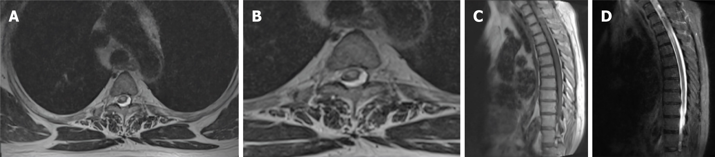
Figure 1 Magnetic resonance imaging of the thoracic spine showing an intra-medullary lesion at the Th1-Th2 level with mixed intensity on T2-weighted.
A and B: Axial sections; C and D: Sagittal sections.
- Citation: Roçi E, Çibuku O, Mara E, Thimjo V, Vyshka G. Incomplete Brown-Séquard syndrome secondary to acenocoumarol-induced hematomyelia: A case report. World J Crit Care Med 2026; 15(3): 117716
- URL: https://www.wjgnet.com/2220-3141/full/v15/i3/117716.htm
- DOI: https://dx.doi.org/10.5492/wjccm.117716