
















Copyright: ©Author(s) 2026.
World J Crit Care Med. Jun 9, 2026; 15(2): 114264
Published online Jun 9, 2026. doi: 10.5492/wjccm.v15.i2.114264
Published online Jun 9, 2026. doi: 10.5492/wjccm.v15.i2.114264
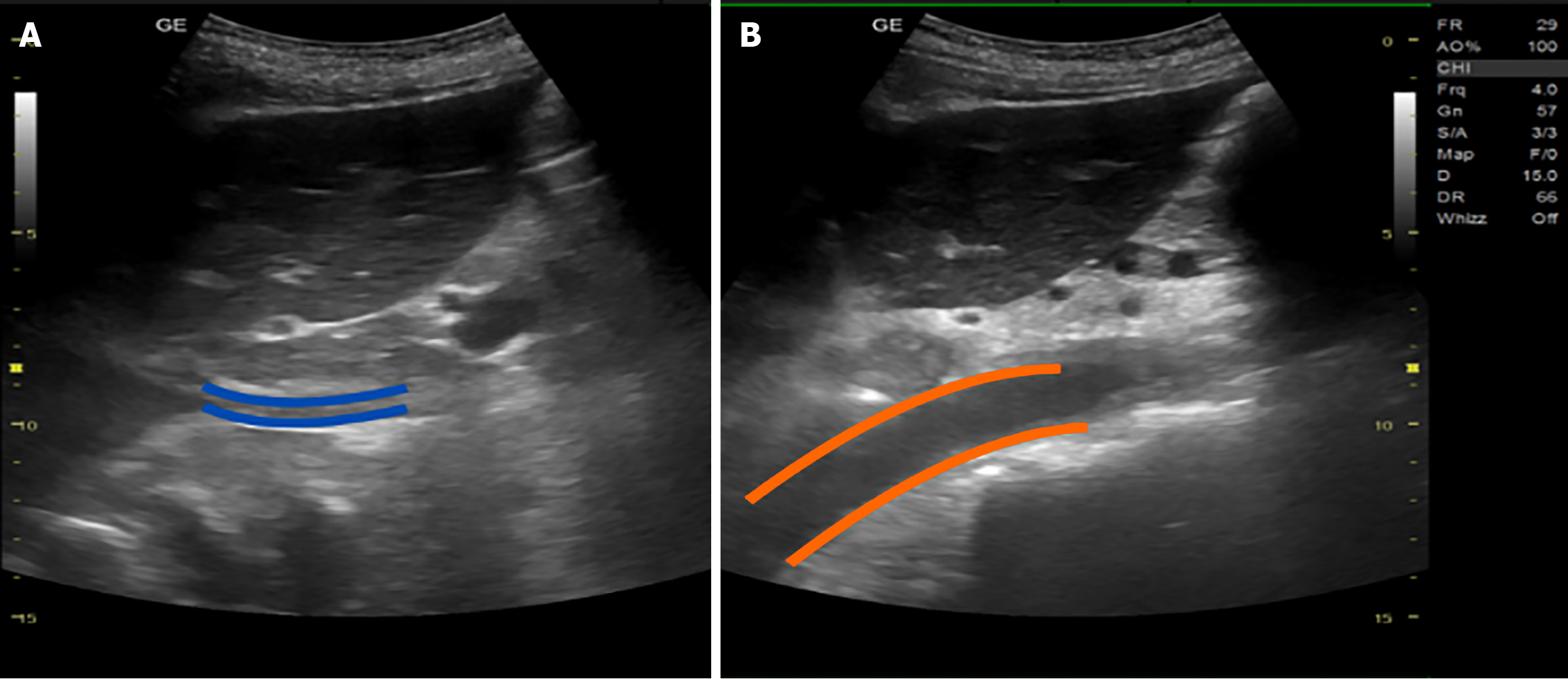
Figure 9 Inferior vena cava (blue marking) and abdominal aorta (orange marking) (longitudinal view).
Note that aorta has a thicker vessel wall and it is more medially located than inferior vena cava (IVC). Blue marking indicates IVC while orange marking indicates abdominal aorta.
- Citation: Lee ZY, Chin WV. Integrating structured point-of-care ultrasound into dengue fever management: A mini review and comprehensive clinical guide. World J Crit Care Med 2026; 15(2): 114264
- URL: https://www.wjgnet.com/2220-3141/full/v15/i2/114264.htm
- DOI: https://dx.doi.org/10.5492/wjccm.v15.i2.114264