
















©The Author(s) 2025.
World J Crit Care Med. Dec 9, 2025; 14(4): 111434
Published online Dec 9, 2025. doi: 10.5492/wjccm.v14.i4.111434
Published online Dec 9, 2025. doi: 10.5492/wjccm.v14.i4.111434
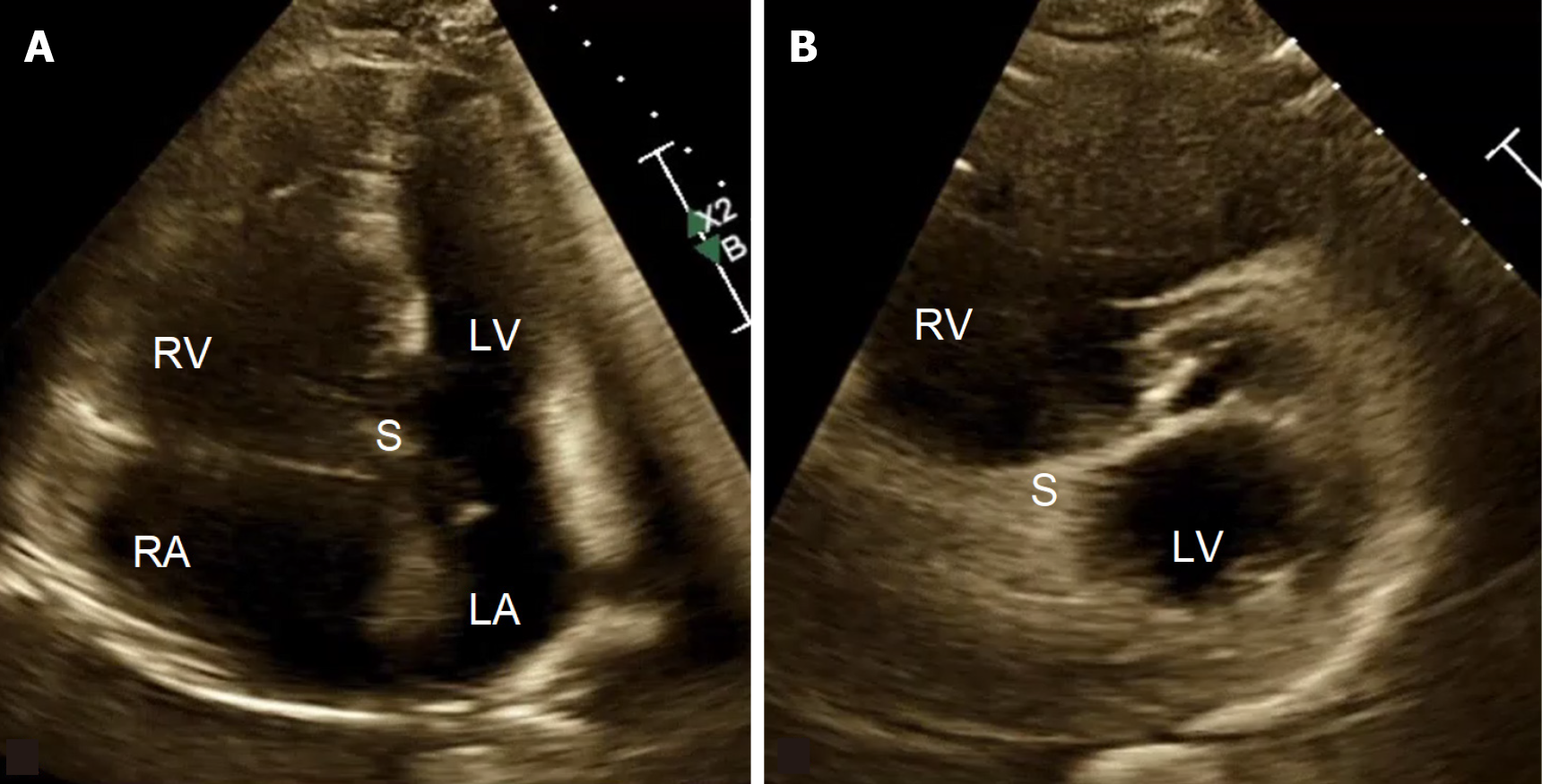
Figure 1 Echocardiographic appearance of right ventricle failure showing RV dilation and compression of the left ventricle, septal flattening, and D-shaping of left ventricle.
A: Apical 4-chamber view; B: Parasternal short-axis view. RV: Right ventricle; LV: Left ventricle; RA: Right atrium; LA: Left atrium; S: Interventricular septum.
- Citation: Kermanian R, Dosanjh H, Lewis MI, Matusov Y. Pathophysiology and management of right ventricular failure in critically ill patients: A narrative review. World J Crit Care Med 2025; 14(4): 111434
- URL: https://www.wjgnet.com/2220-3141/full/v14/i4/111434.htm
- DOI: https://dx.doi.org/10.5492/wjccm.v14.i4.111434