
















©The Author(s) 2025.
World J Crit Care Med. Dec 9, 2025; 14(4): 110669
Published online Dec 9, 2025. doi: 10.5492/wjccm.v14.i4.110669
Published online Dec 9, 2025. doi: 10.5492/wjccm.v14.i4.110669
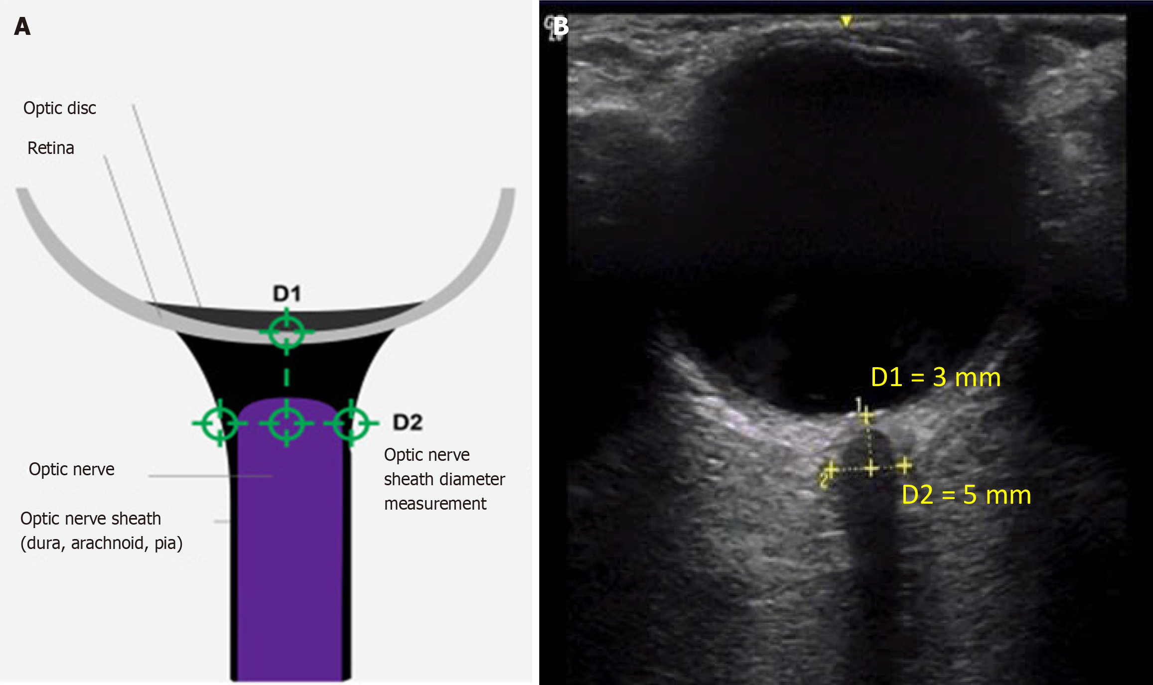
Figure 1 Anatomical landmarks and ultrasonographic measurements of optic nerve sheath diameter.
A: Anatomical landmarks used for optic nerve sheath diameter measurements; B: Enlarged optic nerve sheath on transorbital ultrasonography in a patient with traumatic brain injury. The patient was examined in the supine position with eyes gently closed, and a copious amount of acoustic gel was applied to the upper eyelid to prevent direct pressure on the globe. The probe was positioned lightly over the closed eyelid in the axial plane to visualize the optic nerve posterior to the globe. Two perpendicular diameters of the optic nerve sheath were measured 3 mm posterior to the retina: The vertical diameter (D1 = 3 mm) and the horizontal diameter (D2 = 5 mm). The mean values of D2 were used for further analysis.
- Citation: Truong DMT, Nguyen MHN, Nguyen HQ, Vo LT, Nguyen TT. Optic nerve sheath diameter trajectories and mortality in children with clinically relevant elevated intracranial pressure. World J Crit Care Med 2025; 14(4): 110669
- URL: https://www.wjgnet.com/2220-3141/full/v14/i4/110669.htm
- DOI: https://dx.doi.org/10.5492/wjccm.v14.i4.110669