Copyright
©The Author(s) 2025.
World J Crit Care Med. Sep 9, 2025; 14(3): 101462
Published online Sep 9, 2025. doi: 10.5492/wjccm.v14.i3.101462
Published online Sep 9, 2025. doi: 10.5492/wjccm.v14.i3.101462
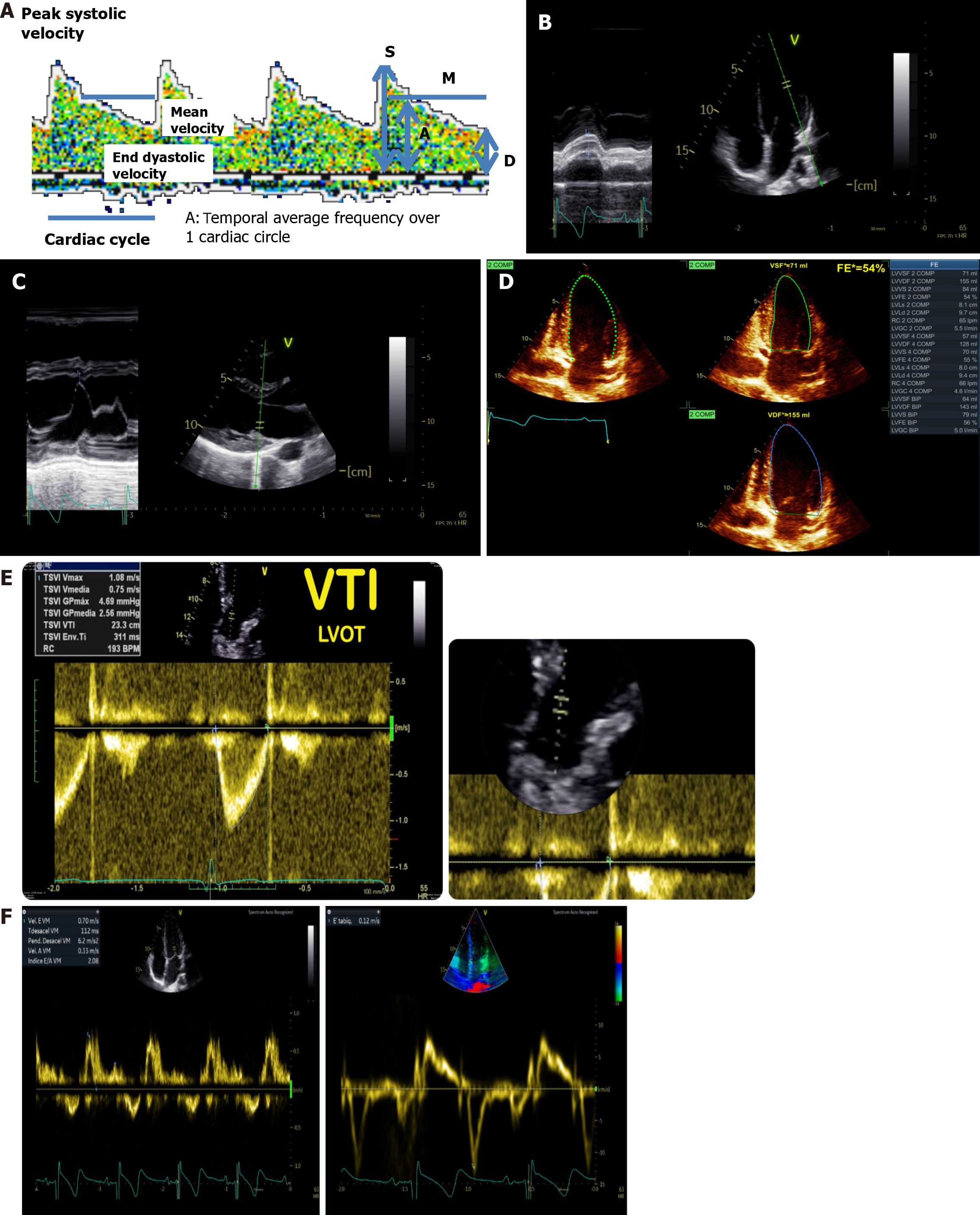
Figure 6 Assessment of cardio-cerebral coupling through ultrasound.
A: Transcranial Doppler spectrum real measurements; B: Mitral annulus plane systolic excursion measurements; C: E point septal separation; D: Ejection fraction by biplane Simpson’s method; E: Left ventricular outflow tract velocity time integral measurement; F: Transmitral Doppler. Left: Normal biphasic morphology, showing distinct E and A waves. Right: Normal triphasic morphology, with the addition of an L wave observed between the E and A waves. LVOT VTI: Left ventricular outflow tract velocity time.
- Citation: Previgliano IJ, Aboumarie HS, Tamagnone FM, Merlo PM, Sosa FA, Feijoo J, Carruega MC. Point of care ultrasound evaluation of cardio-cerebral coupling. World J Crit Care Med 2025; 14(3): 101462
- URL: https://www.wjgnet.com/2220-3141/full/v14/i3/101462.htm
- DOI: https://dx.doi.org/10.5492/wjccm.v14.i3.101462


