Copyright
©The Author(s) 2022.
World J Crit Care Med. Sep 9, 2022; 11(5): 335-341
Published online Sep 9, 2022. doi: 10.5492/wjccm.v11.i5.335
Published online Sep 9, 2022. doi: 10.5492/wjccm.v11.i5.335
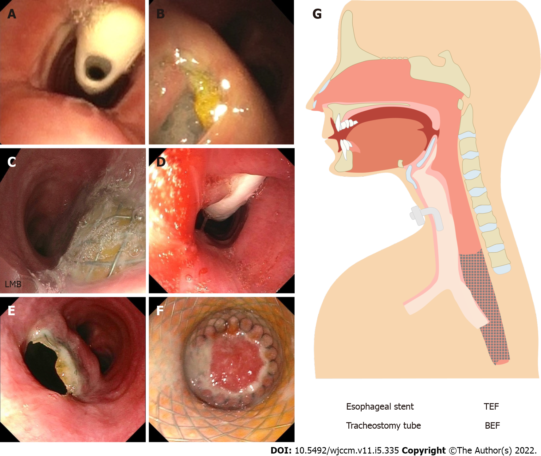
Figure 3 Flexible bronchoscopy at 17 wk.
A: Visualization of tracheostomy tube (asterisk) shortly after bronchoscope is advanced through vocal cords; B: Esophageal lumen visualized at the level of mid-trachea confirming TEF. Flexible Bronchoscopy at 7 wk; C: Protruding esophageal stent through left main bronchi BEF. Esophagoduodenoscopy at 28 wk; D: Visualization of tracheostomy tube (asterisk) through a combined lumen of the esophagus and trachea at 14 cm; E: Proximal end of the esophageal stent located below the end of the tracheotomy at 23cm with a double lumen track, esophagus at 8 o’clock and trachea at 2 o’clock; F: Complete obliteration of esophageal stent due to in-growth of tissue at 35 cm (asterisk); G: Schematic diagram. LMB: Left main bronchi; TEF: Tracheoesophageal fistula; BEF: Bronchoesophageal fistula.
- Citation: Lagrotta G, Ayad M, Butt I, Danckers M. Cardiac arrest due to massive aspiration from a broncho-esophageal fistula: A case report. World J Crit Care Med 2022; 11(5): 335-341
- URL: https://www.wjgnet.com/2220-3141/full/v11/i5/335.htm
- DOI: https://dx.doi.org/10.5492/wjccm.v11.i5.335


