Copyright
©The Author(s) 2018.
World J Crit Care Med. Feb 4, 2018; 7(1): 1-8
Published online Feb 4, 2018. doi: 10.5492/wjccm.v7.i1.1
Published online Feb 4, 2018. doi: 10.5492/wjccm.v7.i1.1
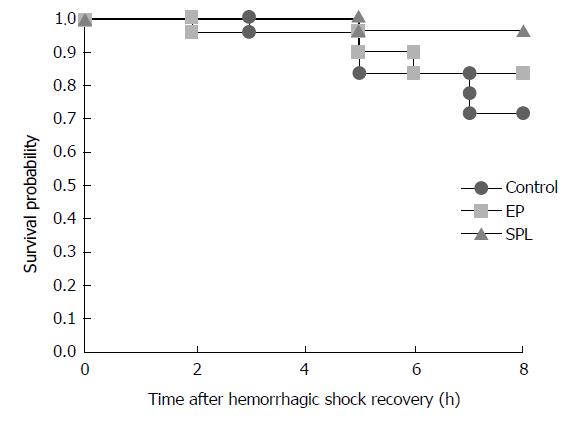
Figure 1 Kaplan–Meier survival analysis.
The survival rates 8 h after recovery from HS were 71%, 94%, and 82% in the control group, SPL group, and EP group, respectively. There were no significant differences in survival rate among the three groups (P = 0.219). HS: Hemorrhagic shock; EP: Eplerenone; SPL: Spironolactone.
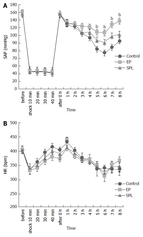
Figure 2 Changes in systolic arterial pressure and heart rate over an 8-h period after hemorrhagic shock recovery.
Changes in (A) SAP and (B) HR in three groups. SAPs gradually decreased after HS recovery in all groups. There were significant difference among three groups in a change of the SAP (P < 0.01). There were significant difference between EP group and control group in SAP at 5-8 h after the HS recovery. EP vs control; P < 0.01(5-8 h). Data represent means ± SE. bP < 0.01 compared with controls. SAP: Systolic arterial pressure; HR: Heart rate; HS: Hemorrhagic shock; EP: Eplerenone.
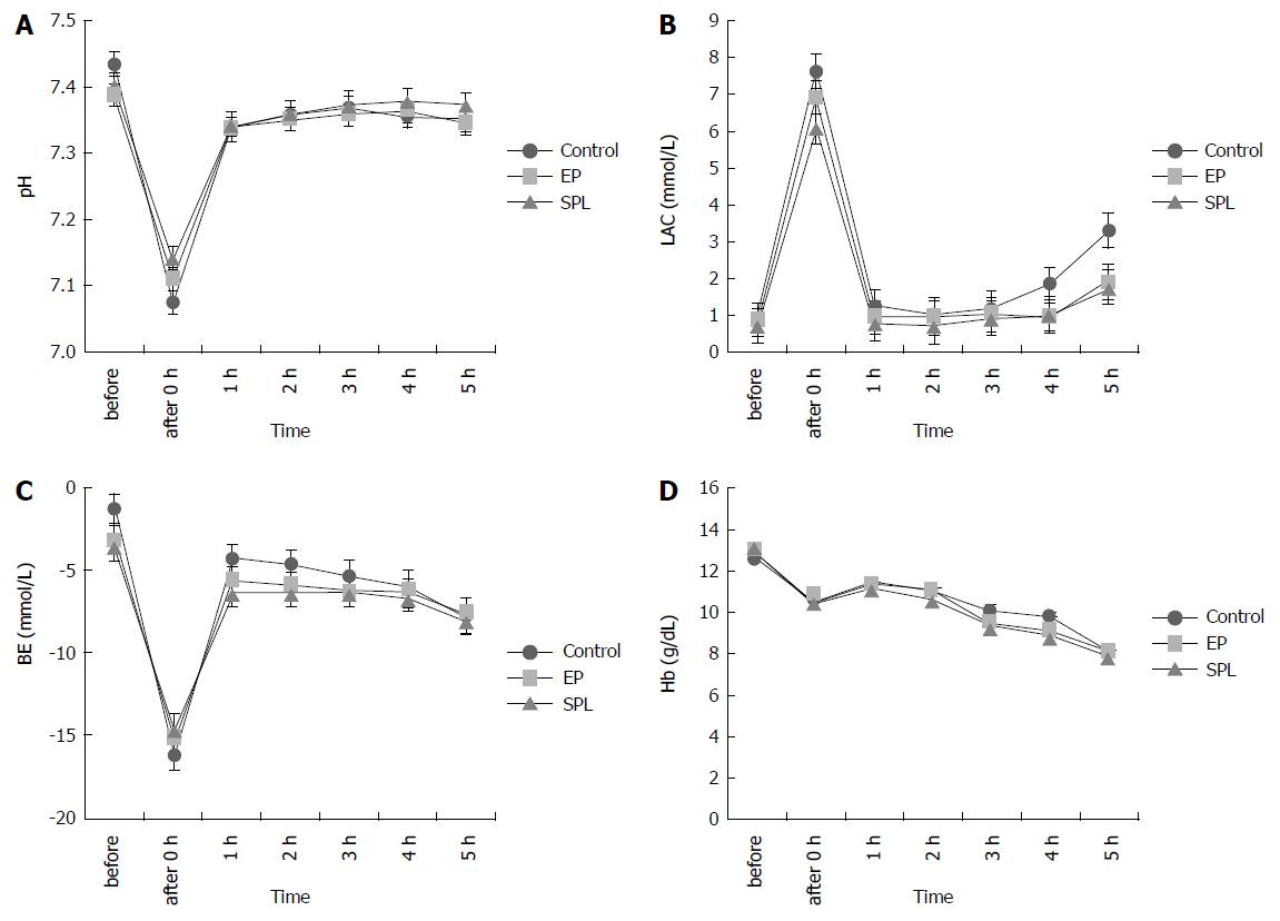
Figure 3 Blood gas analysis results for a 5-h period after hemorrhagic shock recovery.
Changes in (A) PH, (B) LAC, (C) (BE), and (D) Hb in three groups. The lactate concentrations increased immediately after HS, but recovered in all groups. There were no significant differences among the three groups in PH, LAC, BE, and BE. Data represent means ± SE. LAC: Lactic acid; BE: Base excess; Hb: Hemoglobin; HS: Hemorrhagic shock.
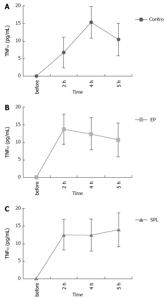
Figure 4 Concentration of plasma tumor necrosis factor-alpha after hemorrhagic shock recovery.
Changes of plasma TNF-α concentration in (A) control, (B) EP and (C) SPL group. The plasma TNF-α concentration in all groups increased slightly after the recovery from HS. There were no significant differences in plasma TNF-α concentrations among the three groups. Data represent means ± SE. SPL: Spironolactone; HS: Hemorrhagic shock; EP: Eplerenone; TNF-α: Tumor necrosis factor-alpha.
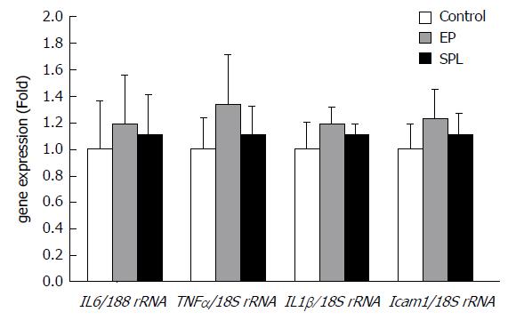
Figure 5 Liver gene expression levels 1 h after hemorrhagic shock recovery.
The relative mRNA expression levels of IL-6/18S rRNA, TNFα/18S rRNA, IL-1β/18S rRNA, and ICAM-1/18S rRNA. These did not exhibit significant differences in the liver at 1 h after hemorrhagic shock recovery among the three groups. Data represent means ± SE.
- Citation: Yamamoto K, Yamamoto T, Takamura M, Usui S, Murai H, Kaneko S, Taniguchi T. Effects of mineralocorticoid receptor antagonists on responses to hemorrhagic shock in rats. World J Crit Care Med 2018; 7(1): 1-8
- URL: https://www.wjgnet.com/2220-3141/full/v7/i1/1.htm
- DOI: https://dx.doi.org/10.5492/wjccm.v7.i1.1


