Copyright
©The Author(s) 2015.
World J Surg Proced. Nov 28, 2015; 5(3): 229-234
Published online Nov 28, 2015. doi: 10.5412/wjsp.v5.i3.229
Published online Nov 28, 2015. doi: 10.5412/wjsp.v5.i3.229
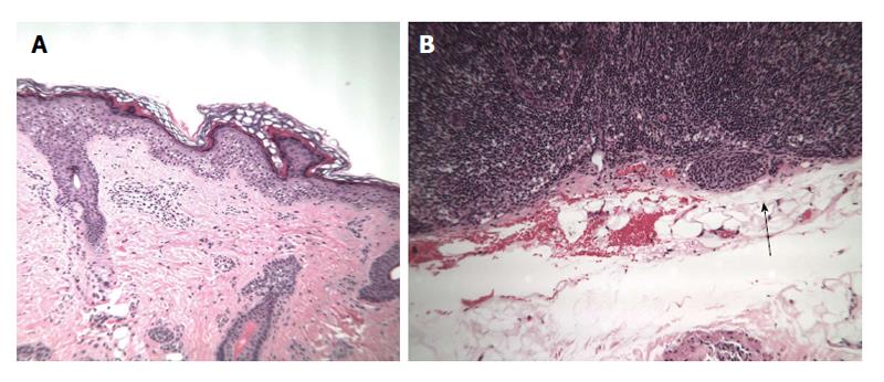
Figure 4 H and E stain.
A: Photomicrograph of primary lesion removed from the left forearm from case 3; B: Photomicrograph of the sentinel lymph node from case 3 - H and E stain - showing subcapsular deposits of pigmented cells (arrow).
- Citation: Psaltis J, Reintgen E, Antar A, Giori M, Alvin L, Benjamin A, Budny B, Gianangelo T, Gruman A, Stamas A, Reintgen M, Giuliano R, Smith J, Reintgen D. Malignant melanoma in the pediatric population. World J Surg Proced 2015; 5(3): 229-234
- URL: https://www.wjgnet.com/2219-2832/full/v5/i3/229.htm
- DOI: https://dx.doi.org/10.5412/wjsp.v5.i3.229


