Copyright
©The Author(s) 2022.
World J Surg Proced. Nov 24, 2022; 12(2): 13-19
Published online Nov 24, 2022. doi: 10.5412/wjsp.v12.i2.13
Published online Nov 24, 2022. doi: 10.5412/wjsp.v12.i2.13
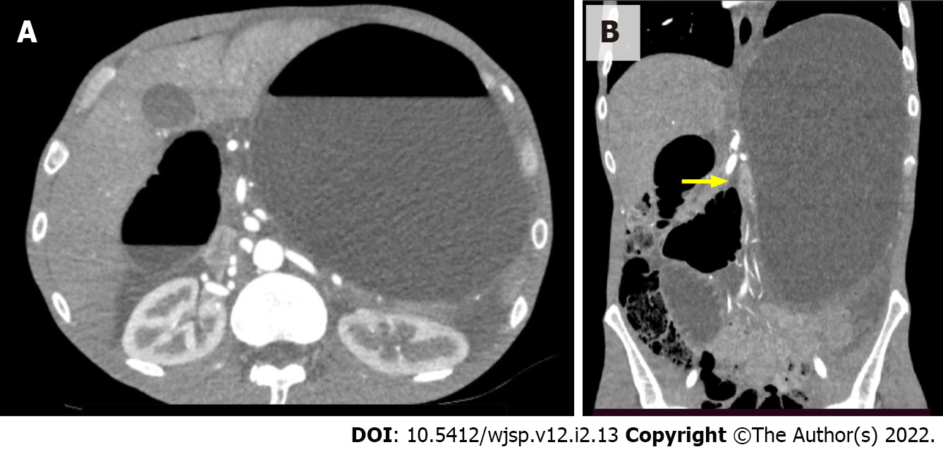
Figure 1 Computed tomography images.
A: Axial computed tomography image showing significant gastric and duodenal distension with formation of air–fluid levels; B: Coronal computed tomography image showing significant gastric and duodenal distension with an abrupt reduction in bowel caliber at the duodenojejunal flexure (yellow arrow).
- Citation: Barros LCTR, Santos BMRTD, Ferreira GSA, Gomes VMDS, Vieira LPB. Superior mesenteric artery syndrome in a patient with esophageal stenosis: A case report. World J Surg Proced 2022; 12(2): 13-19
- URL: https://www.wjgnet.com/2219-2832/full/v12/i2/13.htm
- DOI: https://dx.doi.org/10.5412/wjsp.v12.i2.13


