
















©The Author(s) 2018.
World J Clin Pediatr. Feb 8, 2018; 7(1): 62-66
Published online Feb 8, 2018. doi: 10.5409/wjcp.v7.i1.62
Published online Feb 8, 2018. doi: 10.5409/wjcp.v7.i1.62
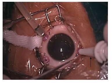
Figure 1 Intraoperative picture of a left eye.
The three sclerotomies 1.5 mm from the limbus are labeled as 1 (inferotemporal port), 2 (superotemporal port) and 3 (superonasal port) of the 27-gauge vitrectomy system (silicon band pieces are not shown in this representative photograph). The white arrow shows the infusion tube through which balanced salt solution flows and maintains intraocular pressure. The black arrow shows the trocar handle inserting the last superonasal port.
- Citation: Shah PK, Prabhu V, Narendran V. Outcomes of transconjuctival sutureless 27-gauge vitrectomy for stage 4 retinopathy of prematurity. World J Clin Pediatr 2018; 7(1): 62-66
- URL: https://www.wjgnet.com/2219-2808/full/v7/i1/62.htm
- DOI: https://dx.doi.org/10.5409/wjcp.v7.i1.62