
















©The Author(s) 2017.
World J Clin Pediatr. Feb 8, 2017; 6(1): 52-59
Published online Feb 8, 2017. doi: 10.5409/wjcp.v6.i1.52
Published online Feb 8, 2017. doi: 10.5409/wjcp.v6.i1.52
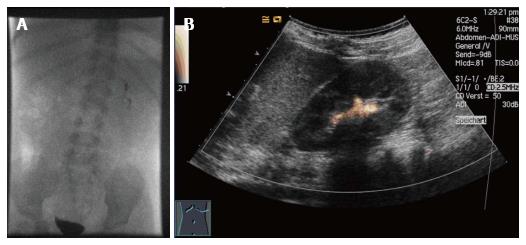
Figure 1 Voiding cystourethrography (A) vs voiding urosonography (B) of a two-year-old male patient with proven vesicoureteral reflux left at the left kidney (grade II).
The voiding urosonography scan shows the urosonography contrast agent microbubbles in the left renal pelvis (agent detection mode).
- Citation: Sauer A, Wirth C, Platzer I, Neubauer H, Veldhoen S, Dierks A, Kaiser R, Kunz A, Beer M, Bley T. Off-label-use of sulfur-hexafluoride in voiding urosonography for diagnosis of vesicoureteral reflux in children: A survey on adverse events. World J Clin Pediatr 2017; 6(1): 52-59
- URL: https://www.wjgnet.com/2219-2808/full/v6/i1/52.htm
- DOI: https://dx.doi.org/10.5409/wjcp.v6.i1.52