
















©The Author(s) 2017.
World J Clin Pediatr. Feb 8, 2017; 6(1): 103-109
Published online Feb 8, 2017. doi: 10.5409/wjcp.v6.i1.103
Published online Feb 8, 2017. doi: 10.5409/wjcp.v6.i1.103
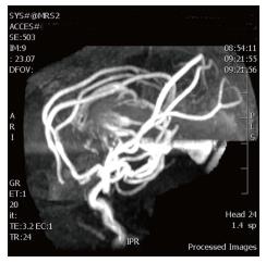
Figure 3 Magnetic resonance angiographic images show multiple enlarged arterial branches from the anterior and posterior cerebral arteries coalescing on the lateral margins of the dilated recipient vein.
- Citation: Puvabanditsin S, Mehta R, Palomares K, Gengel N, Silva CFD, Roychowdhury S, Gupta G, Kashyap A, Sorrentino D. Vein of Galen malformation in a neonate: A case report and review of endovascular management. World J Clin Pediatr 2017; 6(1): 103-109
- URL: https://www.wjgnet.com/2219-2808/full/v6/i1/103.htm
- DOI: https://dx.doi.org/10.5409/wjcp.v6.i1.103