
















©The Author(s) 2017.
World J Clin Pediatr. Feb 8, 2017; 6(1): 10-23
Published online Feb 8, 2017. doi: 10.5409/wjcp.v6.i1.10
Published online Feb 8, 2017. doi: 10.5409/wjcp.v6.i1.10
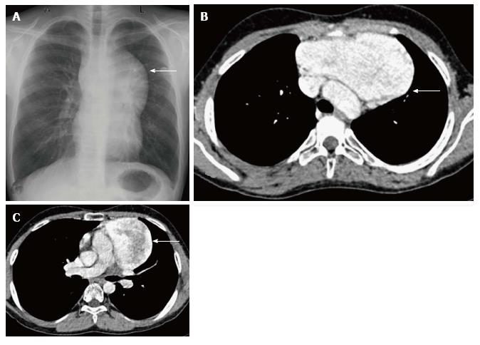
Figure 19 Incidentally detected cavernous hemangioma in 16-year-old male.
Frontal chest radiograph (A) reveals a well-defined, lobulated soft tissue density mass (arrow) with broad base towards the mediastinum and positive hilum overlay sign. CECT axial sections (B and C) show a sharply marginated mass lesion (arrow in B) anterior to the arch of the aorta. Note the intense post contrast enhancement with central heterogeneity (arrow in C).
- Citation: Manchanda S, Bhalla AS, Jana M, Gupta AK. Imaging of the pediatric thymus: Clinicoradiologic approach. World J Clin Pediatr 2017; 6(1): 10-23
- URL: https://www.wjgnet.com/2219-2808/full/v6/i1/10.htm
- DOI: https://dx.doi.org/10.5409/wjcp.v6.i1.10