Copyright
©The Author(s) 2017.
World J Clin Pediatr. Feb 8, 2017; 6(1): 10-23
Published online Feb 8, 2017. doi: 10.5409/wjcp.v6.i1.10
Published online Feb 8, 2017. doi: 10.5409/wjcp.v6.i1.10
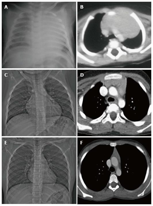
Figure 6 Normal variation in the appearance of the thymus with age.
Frontal radiographs and CECT axial images at ages of (A, B) 2 mo, (C, D) 7 years and (E, F) 12 years. Note that the initial prominent bilobed thymus in a neonate assumes a quadrilateral shape with convex margins in early childhood. Gradually it assumes a triangular configuration with straight margins. The CT density also decreases with age with fatty replacement occurring in adults.
- Citation: Manchanda S, Bhalla AS, Jana M, Gupta AK. Imaging of the pediatric thymus: Clinicoradiologic approach. World J Clin Pediatr 2017; 6(1): 10-23
- URL: https://www.wjgnet.com/2219-2808/full/v6/i1/10.htm
- DOI: https://dx.doi.org/10.5409/wjcp.v6.i1.10


