
















©The Author(s) 2017.
World J Clin Pediatr. Feb 8, 2017; 6(1): 10-23
Published online Feb 8, 2017. doi: 10.5409/wjcp.v6.i1.10
Published online Feb 8, 2017. doi: 10.5409/wjcp.v6.i1.10
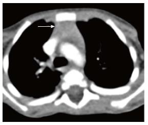
Figure 5 Normal appearance of the thymus on computed tomography scan of a 3-year-old boy.
Axial contrast-enhanced computed tomography section shows homogenous soft tissue density structure (arrow) in the anterior mediastinum. Note that there is no compression of the vascular structures or airway.
- Citation: Manchanda S, Bhalla AS, Jana M, Gupta AK. Imaging of the pediatric thymus: Clinicoradiologic approach. World J Clin Pediatr 2017; 6(1): 10-23
- URL: https://www.wjgnet.com/2219-2808/full/v6/i1/10.htm
- DOI: https://dx.doi.org/10.5409/wjcp.v6.i1.10