Copyright
©The Author(s) 2016.
World J Clin Pediatr. Feb 8, 2016; 5(1): 35-46
Published online Feb 8, 2016. doi: 10.5409/wjcp.v5.i1.35
Published online Feb 8, 2016. doi: 10.5409/wjcp.v5.i1.35
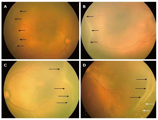
Figure 1 RetCam fundus images showing retinopathy of prematurity stages 1, 2, 3 and 4A.
A: Fundus image of right eye showing stage 1 ROP with demarcation line (black arrows); B: Fundus image of right eye showing stage 2 ROP with ridge (black arrows); C: Fundus image of left eye showing stage 3 extra retinal fibrovascular proliferation (black arrows); D: Fundus picture of left eye showing stage 4A partial retinal detachment not involving the fovea (black arrows). Laser scars are shown with white arrows. ROP: Retinopathy of prematurity.
- Citation: Shah PK, Prabhu V, Karandikar SS, Ranjan R, Narendran V, Kalpana N. Retinopathy of prematurity: Past, present and future. World J Clin Pediatr 2016; 5(1): 35-46
- URL: https://www.wjgnet.com/2219-2808/full/v5/i1/35.htm
- DOI: https://dx.doi.org/10.5409/wjcp.v5.i1.35


