Copyright
©2013 Baishideng Publishing Group Co.
World J Clin Pediatr. Nov 8, 2013; 2(4): 54-64
Published online Nov 8, 2013. doi: 10.5409/wjcp.v2.i4.54
Published online Nov 8, 2013. doi: 10.5409/wjcp.v2.i4.54
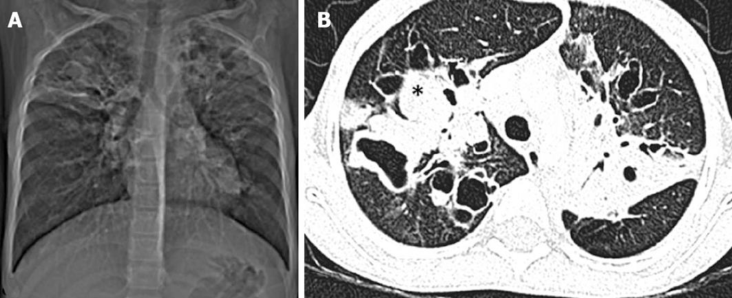
Figure 7 A 7-year-old boy with cystic fibrosis.
Computed tomography (CT) scout image (A) and axial CT chest image (B) showing bilateral upper lobe bronchiectasis with bronchocele formation (asterisk) due to mucous plugging and sparing of lower zones.
- Citation: Singh D, Bhalla AS, Veedu PT, Arora A. Imaging evaluation of hemoptysis in children. World J Clin Pediatr 2013; 2(4): 54-64
- URL: https://www.wjgnet.com/2219-2808/full/v2/i4/54.htm
- DOI: https://dx.doi.org/10.5409/wjcp.v2.i4.54


