
















Copyright: ©Author(s) 2026.
World J Clin Pediatr. Dec 9, 2026; 15(4): 120972
Published online Dec 9, 2026. doi: 10.5409/wjcp.120972
Published online Dec 9, 2026. doi: 10.5409/wjcp.120972
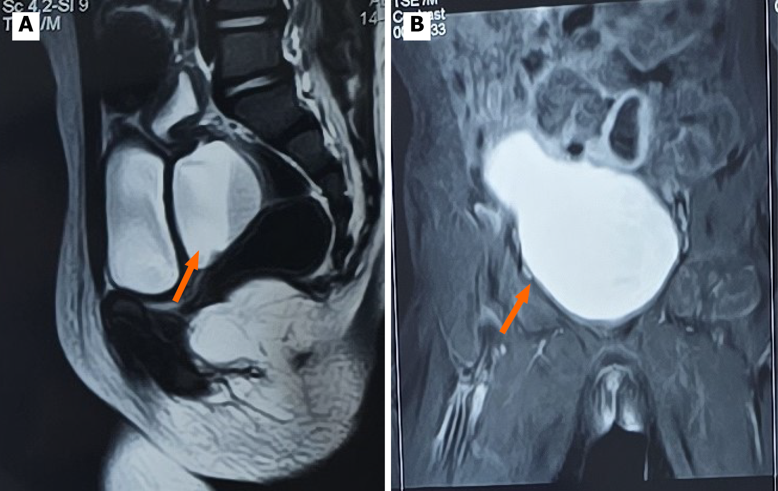
Figure 1 Magnetic resonance imaging.
A: Magnetic resonance imaging T2 W midline sagittal image showing pyometrocolpos (orange arrow); B: Coronal T1 post contrast fat saturated sequence demonstrates enhancing pyometrocolpos (orange arrow).
- Citation: Kanneganti P, Verma A, Upadhyaya VD, Gautam AD. Clinical conundrum in the management of complex urogenital anomaly in a child: A case report. World J Clin Pediatr 2026; 15(4): 120972
- URL: https://www.wjgnet.com/2219-2808/full/v15/i4/120972.htm
- DOI: https://dx.doi.org/10.5409/wjcp.120972