
















Copyright: ©Author(s) 2026.
World J Clin Pediatr. Dec 9, 2026; 15(4): 120132
Published online Dec 9, 2026. doi: 10.5409/wjcp.120132
Published online Dec 9, 2026. doi: 10.5409/wjcp.120132
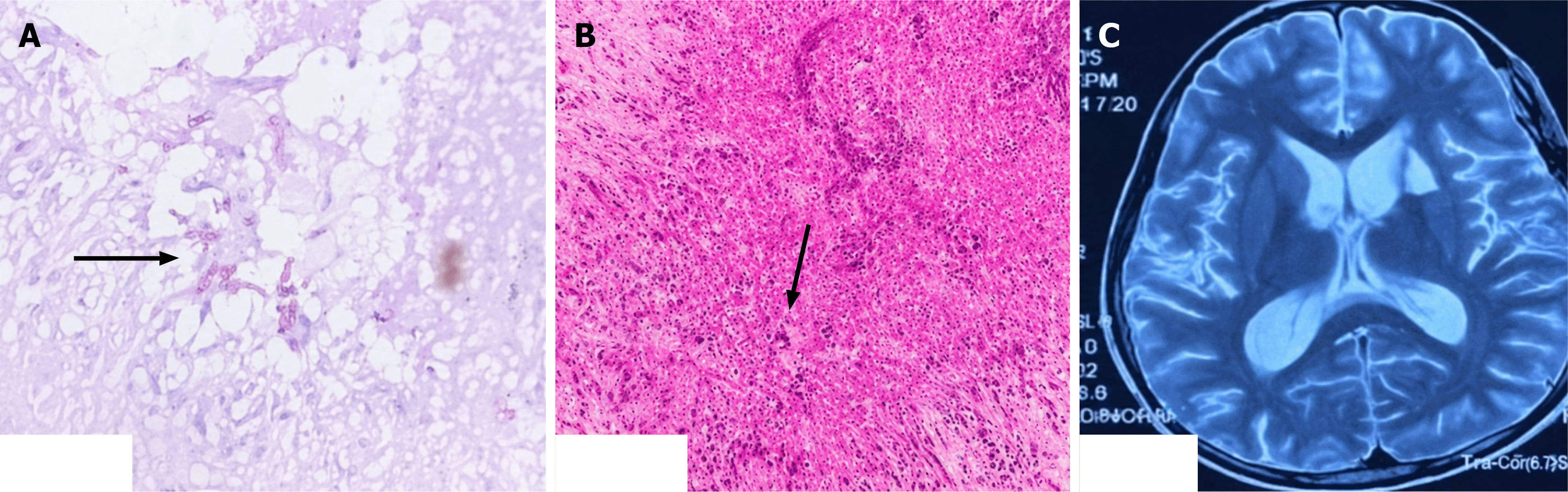
Figure 2 Microscopy of abscess material and a post therapy repeat magnetic resonance imaging brain.
A: Pigmented fungal hyphae on direct microscopy of aspirated pus (arrow); B: Necrotizing granulomatous inflammation with central necrosis with multinucleate giant cell (arrow); C: Follow up contrast-enhanced magnetic resonance imaging brain after 6 weeks of amphotericin B showing resolution of abscess.
- Citation: Soni A, Yamala N, Krishnan G, Attilli H, Mittal H. Coexisting phaeohyphomycosis and tubercular central nervous system infections in an immunocompetent child: A case report. World J Clin Pediatr 2026; 15(4): 120132
- URL: https://www.wjgnet.com/2219-2808/full/v15/i4/120132.htm
- DOI: https://dx.doi.org/10.5409/wjcp.120132