
















Copyright: ©Author(s) 2026.
World J Clin Pediatr. Sep 9, 2026; 15(3): 119602
Published online Sep 9, 2026. doi: 10.5409/wjcp.119602
Published online Sep 9, 2026. doi: 10.5409/wjcp.119602
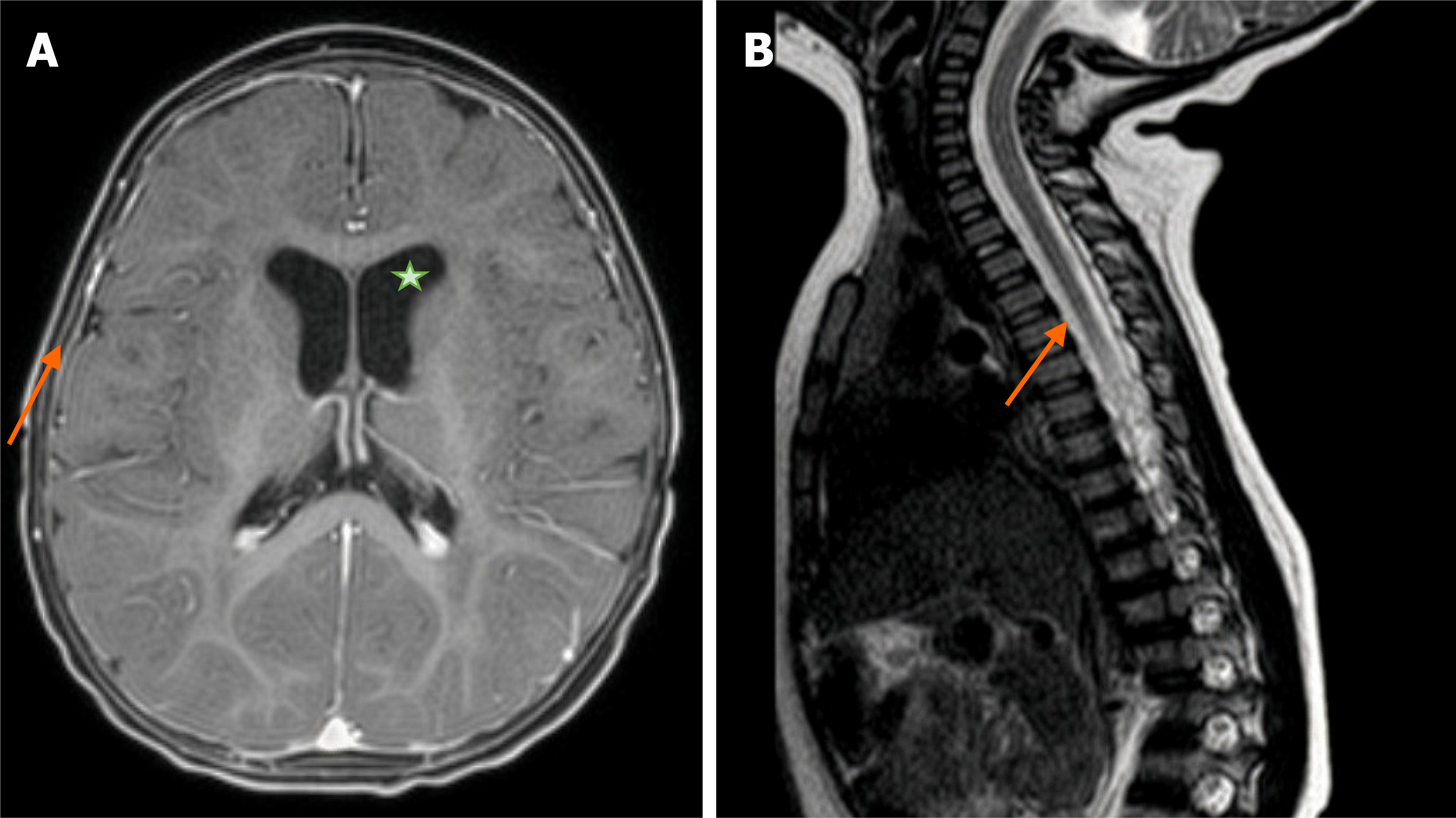
Figure 3 Follow-up magnetic resonance imaging findings.
A: Brain magnetic resonance imaging (MRI) at 17 months of age showing marked reduction in lateral ventricular size (8 mm vs 12 mm at admission), complete resolution of the subdural hygroma, and normal brain parenchyma with preserved cortical sulci. Ventricular enlargement is indicated by an asterisk; B: Cervical spine MRI demonstrating normal spinal cord signal from C3 to C6 without evidence of cord atrophy. The orange arrows indicate the lesion locations in the brain and cervical spine.
- Citation: Nguyen BT, Vo LT, Pham UH, Tran HN, Dang OHT, Nguyen DT, Tran TTH, To LT, Do VC, Nguyen TT. Clinical recovery of severe infantile Angiostrongylus cantonensis meningoencephalomyelitis treated with pulse methylprednisolone: A case report and review of literature. World J Clin Pediatr 2026; 15(3): 119602
- URL: https://www.wjgnet.com/2219-2808/full/v15/i3/119602.htm
- DOI: https://dx.doi.org/10.5409/wjcp.119602