
















Copyright: ©Author(s) 2026.
World J Clin Pediatr. Sep 9, 2026; 15(3): 119602
Published online Sep 9, 2026. doi: 10.5409/wjcp.119602
Published online Sep 9, 2026. doi: 10.5409/wjcp.119602
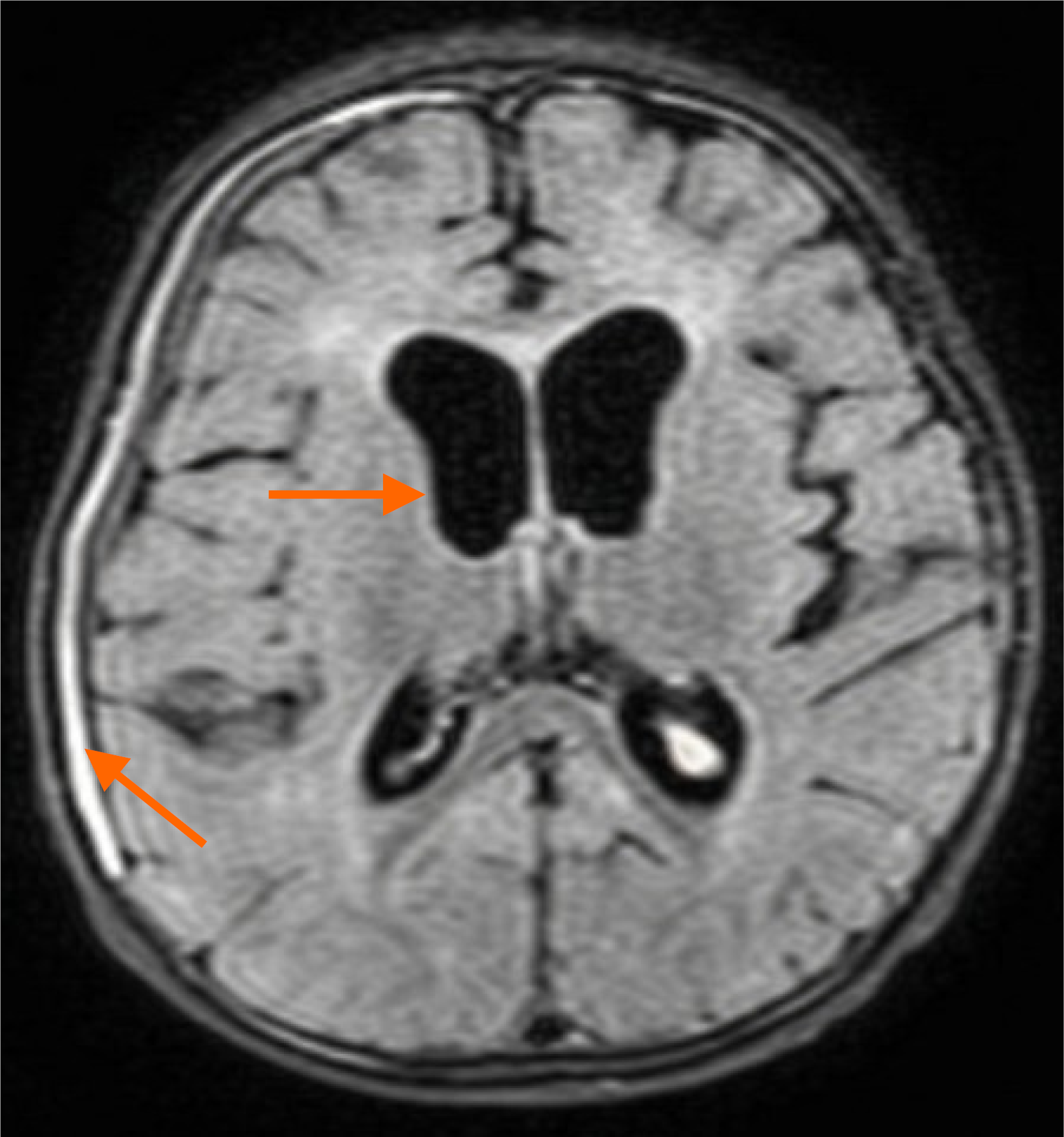
Figure 2 Pre-discharge and follow-up magnetic resonance imaging findings.
Pre-discharge brain magnetic resonance imaging demonstrating a thin right-sided subdural hygroma along the cerebral convexity and persistent ventriculomegaly as residual treatment-related sequelae. The orange arrows indicate the lesion locations in the brain.
- Citation: Nguyen BT, Vo LT, Pham UH, Tran HN, Dang OHT, Nguyen DT, Tran TTH, To LT, Do VC, Nguyen TT. Clinical recovery of severe infantile Angiostrongylus cantonensis meningoencephalomyelitis treated with pulse methylprednisolone: A case report and review of literature. World J Clin Pediatr 2026; 15(3): 119602
- URL: https://www.wjgnet.com/2219-2808/full/v15/i3/119602.htm
- DOI: https://dx.doi.org/10.5409/wjcp.119602