
















Copyright: ©Author(s) 2026.
World J Clin Pediatr. Sep 9, 2026; 15(3): 119602
Published online Sep 9, 2026. doi: 10.5409/wjcp.119602
Published online Sep 9, 2026. doi: 10.5409/wjcp.119602
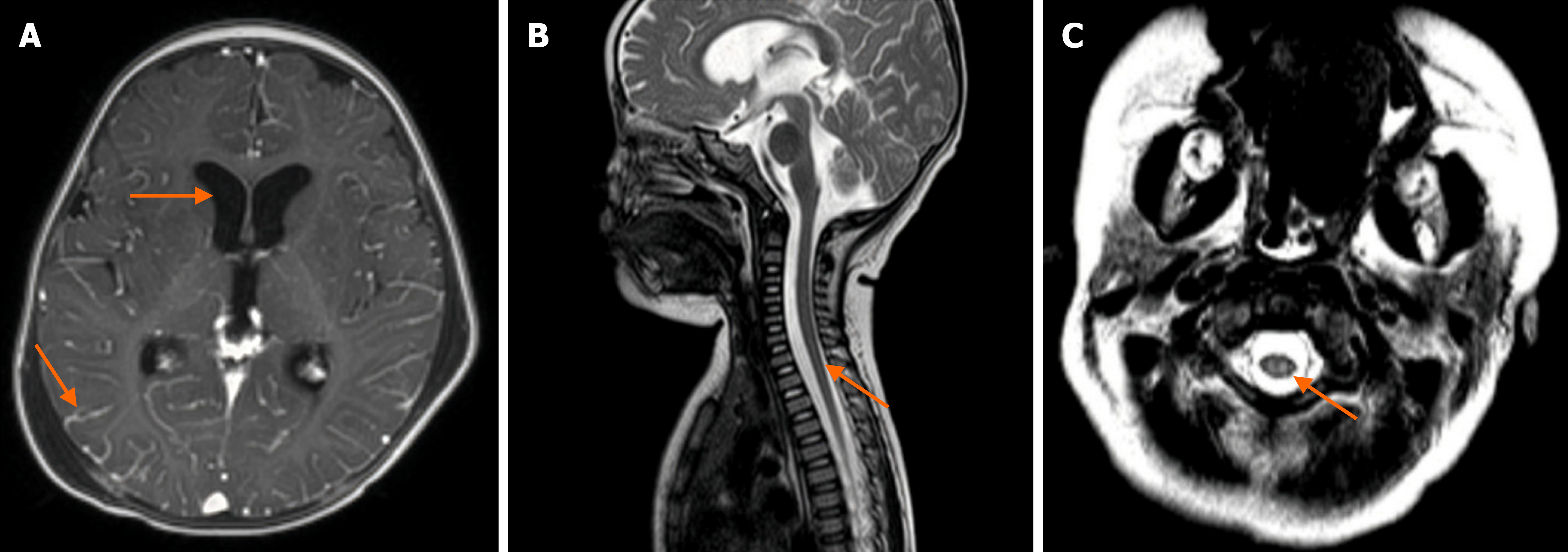
Figure 1 Initial neuroimaging findings.
A: Axial T2-weighted brain magnetic resonance imaging showing diffuse bilateral hyperintense lesions involving the gray and white matter of the frontal, parietal, and occipital lobes with associated ventricular dilatation. No acute hemorrhage, mass effect, or significant ventri
- Citation: Nguyen BT, Vo LT, Pham UH, Tran HN, Dang OHT, Nguyen DT, Tran TTH, To LT, Do VC, Nguyen TT. Clinical recovery of severe infantile Angiostrongylus cantonensis meningoencephalomyelitis treated with pulse methylprednisolone: A case report and review of literature. World J Clin Pediatr 2026; 15(3): 119602
- URL: https://www.wjgnet.com/2219-2808/full/v15/i3/119602.htm
- DOI: https://dx.doi.org/10.5409/wjcp.119602