
















Copyright: ©Author(s) 2026.
World J Clin Pediatr. Sep 9, 2026; 15(3): 119506
Published online Sep 9, 2026. doi: 10.5409/wjcp.119506
Published online Sep 9, 2026. doi: 10.5409/wjcp.119506
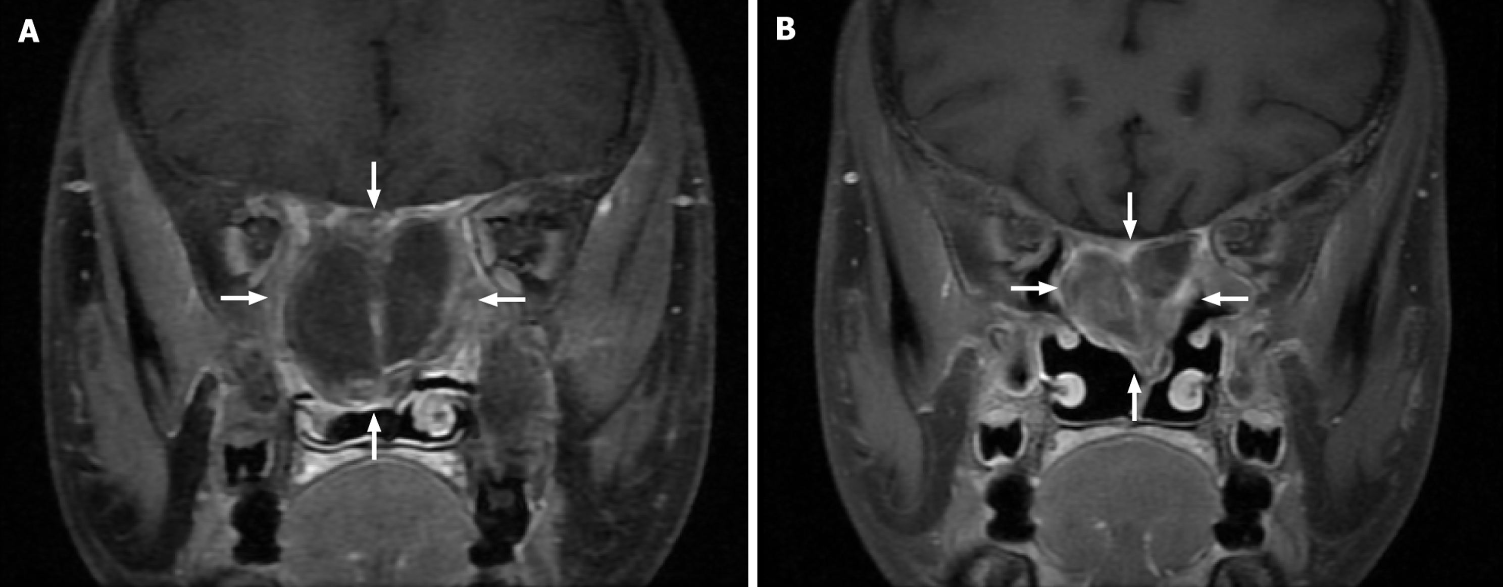
Figure 2 Magnetic resonance imaging of a patient with composite hemangioendothelioma before and after sirolimus therapy.
A: Baseline post-contrast T1-weighted image with fat suppression in the coronal view demonstrating a large heterogeneous mass with areas of necrosis involving the nasal cavity, sphenoid sinuses, and ethmoid sinuses (arrows); B: Follow-up magnetic resonance imaging obtained after 6 months of sirolimus therapy showing a marked reduction in tumor size (arrows).
- Citation: Thongngam N, Sathitsamitphong L, Morakote W, Chanthong S, Maneekesorn S, Natesirinilkul R, Charoenkwan P, Choed-Amphai C. Sirolimus for the treatment of vascular tumors and malformations in children: A 10-year single-institution retrospective study. World J Clin Pediatr 2026; 15(3): 119506
- URL: https://www.wjgnet.com/2219-2808/full/v15/i3/119506.htm
- DOI: https://dx.doi.org/10.5409/wjcp.119506