
















Copyright: ©Author(s) 2026.
World J Clin Pediatr. Sep 9, 2026; 15(3): 117421
Published online Sep 9, 2026. doi: 10.5409/wjcp.117421
Published online Sep 9, 2026. doi: 10.5409/wjcp.117421
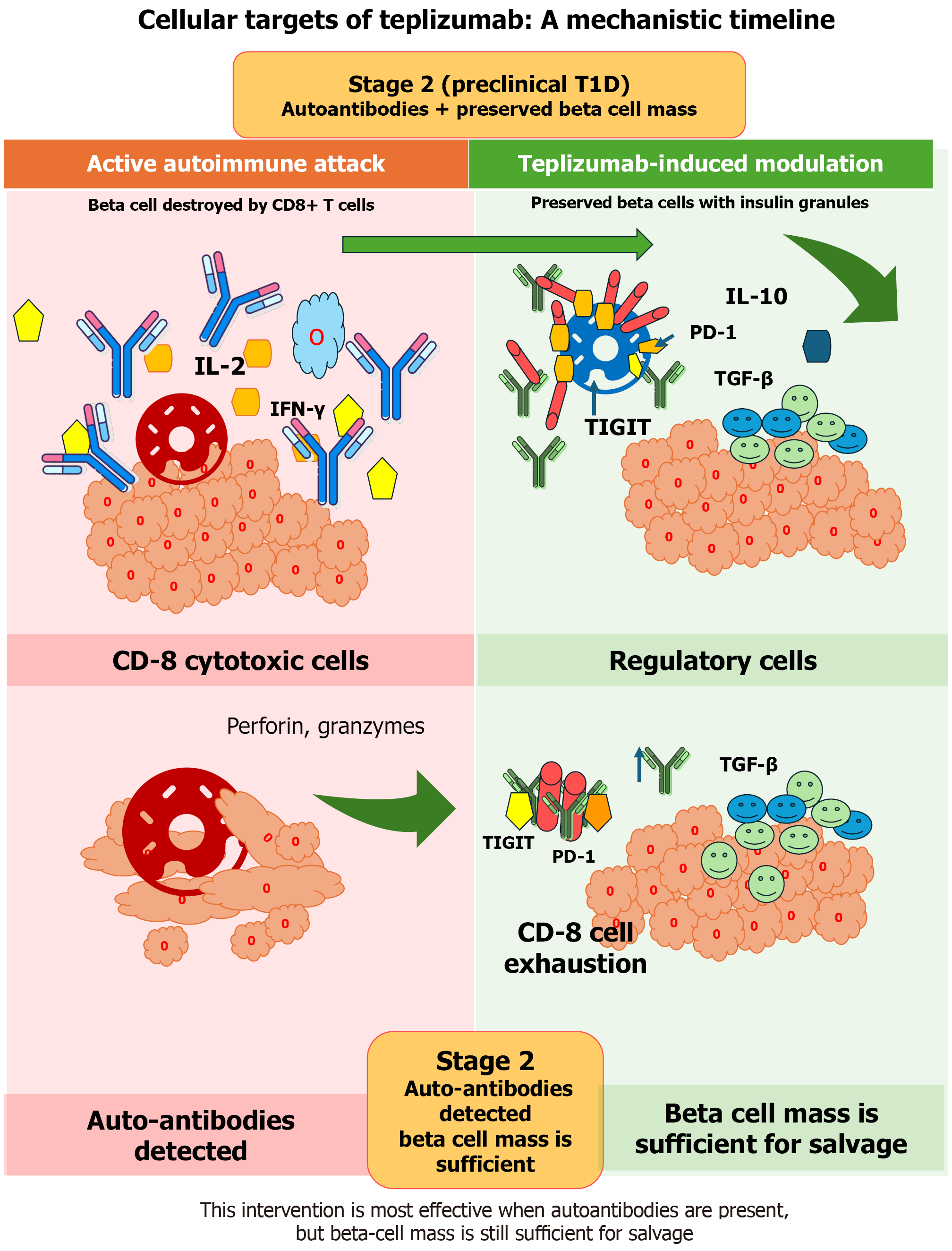
Figure 11 Mechanistic action of teplizumab and β-cell preservation in stage 2 type 1 diabetes.
This figure illustrates the cellular and immunological mechanisms by which teplizumab delays disease progression in stage 2 (preclinical) type 1 diabetes, highlighting the critical importance of treatment timing. The schematic is presented as a before-after comparison. On the left, an active autoimmune attack is shown, characterized by autoreactive CD8⁺ cytotoxic T cells infiltrating the pancreatic islet and inducing β-cell destruction through perforin- and granzyme-mediated cytotoxicity, accompanied by pro-inflammatory cytokine release (e.g., interleukin-2, interferon-gamma). On the right, teplizumab-induced immune modulation is depicted. Teplizumab, an anti-CD3 monoclonal antibody, binds to the CD3ε chain of the T-cell receptor complex on autoreactive CD8⁺ T cells, inducing a metabolic and functional shift toward T-cell “exhaustion” or anergy. This state is characterized by reduced effector function and upregulation of inhibitory markers such as programmed cell death protein 1 and T cell immunoreceptor with immunoglobulin and immunoglobulin and tyrosine-based inhibitory motif domain, resulting in diminished cytokine secretion and cytotoxic activity. Concurrently, teplizumab promotes a regulatory immune network, supporting the expansion and functional activity of regulatory T cells. These cells secrete immunosuppressive cytokines (e.g., interleukin-10 and transforming growth factor-beta), actively suppressing ongoing autoimmune inflammation within the islet microenvironment. The combined effects of effector T-cell attenuation and enhanced immune regulation lead to preservation of residual β-cell mass, maintenance of insulin granules, and prolonged endogenous insulin secretion. A highlighted timeline emphasizes the stage 2 clinical window, defined by the presence of diabetes-associated autoantibodies with preserved β-cell function and normoglycemia. Intervention during this phase maximizes the disease-modifying potential of teplizumab, delaying progression to symptomatic (stage 3) type 1 diabetes. T1D: Type 1 diabetes; IL: Interleukin; IFN: Interferon; PD-1: Programmed cell death protein 1; TGF: Trans
- Citation: Al-Beltagi M, Saeed NK, Bediwy AS, Bediwy EA, Elbeltagi R. From genes to environment: A life-course approach to prevent pediatric autoimmune diseases. World J Clin Pediatr 2026; 15(3): 117421
- URL: https://www.wjgnet.com/2219-2808/full/v15/i3/117421.htm
- DOI: https://dx.doi.org/10.5409/wjcp.117421