
















Copyright: ©Author(s) 2026.
World J Clin Pediatr. Jun 9, 2026; 15(2): 117843
Published online Jun 9, 2026. doi: 10.5409/wjcp.v15.i2.117843
Published online Jun 9, 2026. doi: 10.5409/wjcp.v15.i2.117843
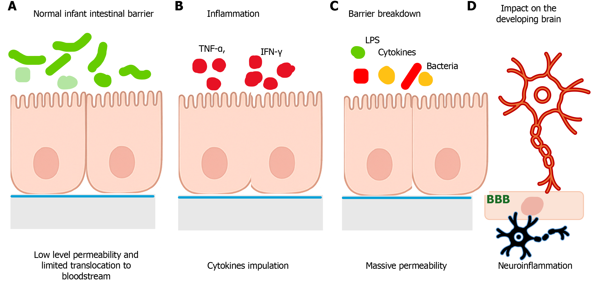
Figure 4 Early-life intestinal barrier integrity and its disruption during inflammation: Pathways linking the gut to the developing brain.
This schematic illustrates the developmental characteristics of the infant intestinal barrier and the mechanisms by which early-life inflammation disrupts gut integrity and triggers downstream neuroinflammatory effects. A: It depicts the normal neonatal intestinal barrier, characterized by immature yet functionally regulated permeability, a thin mucus layer, and developing tight junctions that allow controlled antigen exposure essential for immune maturation; B: It shows inflammatory stimulation by cytokines such as tumor necrosis factor-alpha and interferon gamma, which initiate epithelial stress responses; C: It demonstrates inflammatory breakdown of the barrier: Tight junction disassembly, widening of the paracellular space, and translocation of luminal microbial components – including lipopolysaccharide – along with pro-inflammatory cytokines into the systemic circulation; D: It illustrates how these circulating mediators cross or signal across the immature blood-brain barrier, activate microglia, and induce neuroinflammation, ultimately altering neurodevelopmental processes such as synaptogenesis, neuronal connectivity, and cognitive maturation. Together, it summarizes the mechanistic cascade linking early gastrointestinal inflammation to potential long-term cognitive and behavioral consequences. BBB: Blood-brain barrier; LPS: Lipopolysaccharide.
- Citation: Al-Beltagi M, Saeed NK, El-Sawaf Y, Bediwy AS, Elbeltagi R. Early-life gastrointestinal inflammation and the developing brain: Unravelling the pathways to long-term cognitive dysfunction. World J Clin Pediatr 2026; 15(2): 117843
- URL: https://www.wjgnet.com/2219-2808/full/v15/i2/117843.htm
- DOI: https://dx.doi.org/10.5409/wjcp.v15.i2.117843