
















Copyright: ©Author(s) 2026.
World J Clin Pediatr. Jun 9, 2026; 15(2): 117629
Published online Jun 9, 2026. doi: 10.5409/wjcp.v15.i2.117629
Published online Jun 9, 2026. doi: 10.5409/wjcp.v15.i2.117629
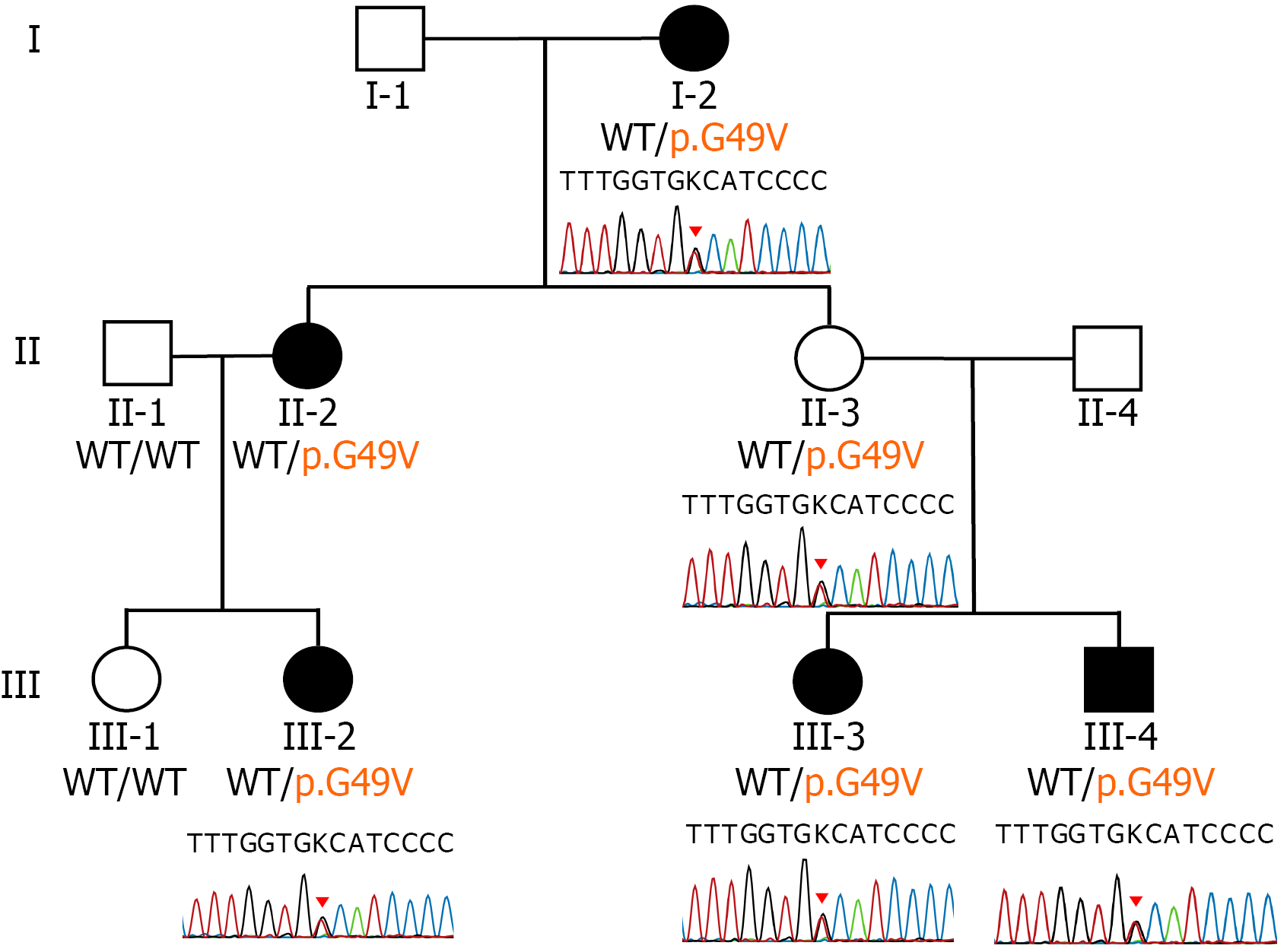
Figure 1 Sanger sequencing electropherograms demonstrating segregation of the novel TMEM63A variant in the family.
Electropherograms showing the heterozygous TMEM63A c.146G>T (p.Gly49Val) variant identified in affected individuals (III-2, III-3, III-4) and carrier relatives (I-2, II-2, II-3). The arrow indicates the position of the nucleotide substitution. Wild-type sequence is shown for comparison. The color version of this figure is available in the online edition. c: Coding DNA sequence position; p: Protein sequence position; WT: Wild type.
- Citation: Chanvanichtrakool M, Kulsirichawaroj P, Jaito W, Pho-Iam T, Kamolvisit W, Likasitwattanakul S. Novel TMEM63A mutation associated with transient hypomyelination of infancy - lessons from a previously negative whole-exome sequencing case: Three case reports. World J Clin Pediatr 2026; 15(2): 117629
- URL: https://www.wjgnet.com/2219-2808/full/v15/i2/117629.htm
- DOI: https://dx.doi.org/10.5409/wjcp.v15.i2.117629