
















Copyright: ©Author(s) 2026.
World J Clin Pediatr. Jun 9, 2026; 15(2): 116956
Published online Jun 9, 2026. doi: 10.5409/wjcp.v15.i2.116956
Published online Jun 9, 2026. doi: 10.5409/wjcp.v15.i2.116956
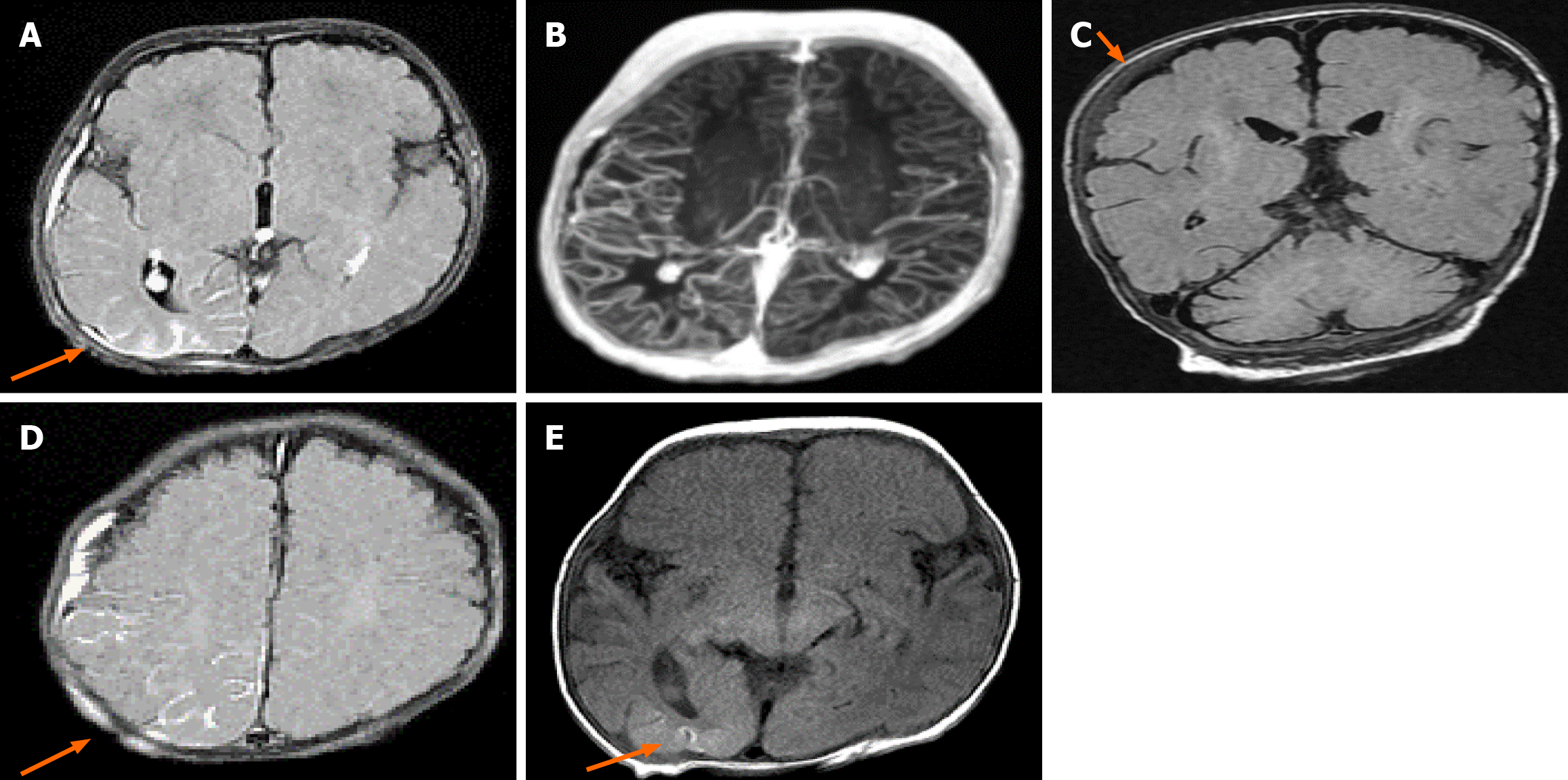
Figure 3 Brain magnetic resonance imaging depicting several abnormalities.
A: Abnormal sulcal hyperintensity (‘ivy sign’) leptomeningeal enhancement over the right parieto-occipital lobe and peri-rolandic region (arrow); B: Diffuse tortuosity of the intracranial arteries on axial T2 image and post-contrast axial T1-weighted maximum intensity projection image; C: Hyperintense 3 mm subdural hematoma on the right frontal lobe on coronal fluid-attenuated inversion recovery image (arrow); D: Contrast-enhancement within the tabula of the right parietal bone (arrow); E: Hyperintense laminar cortical necrosis of right occipital lobe (arrow).
- Citation: Hadi Y, Khawaja N, Ben Turkia H, Nasef M, Baili S. Prolonged survival in Menkes disease with a novel ATP7A variant: A case report. World J Clin Pediatr 2026; 15(2): 116956
- URL: https://www.wjgnet.com/2219-2808/full/v15/i2/116956.htm
- DOI: https://dx.doi.org/10.5409/wjcp.v15.i2.116956