
















Copyright: ©Author(s) 2026.
World J Clin Pediatr. Jun 9, 2026; 15(2): 116098
Published online Jun 9, 2026. doi: 10.5409/wjcp.v15.i2.116098
Published online Jun 9, 2026. doi: 10.5409/wjcp.v15.i2.116098
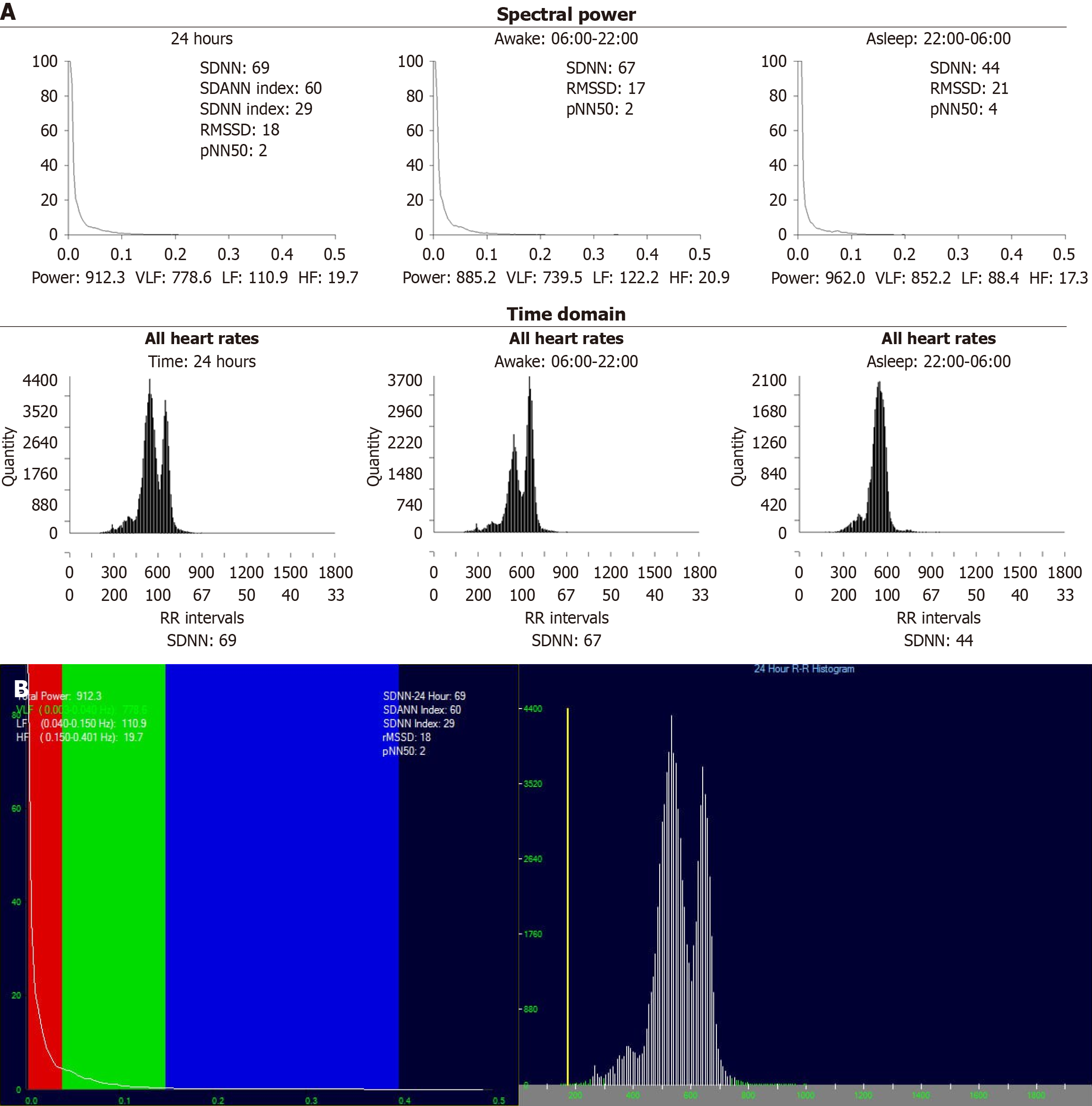
Figure 1 Heart rate variability analysis in a thalassemia patient.
A: Spectral and time-domain plots of heart rate variability (HRV) in an 8-year-old child with β-thalassemia major over 24 hours, showing both awake (06:00-22:00) and sleep (22:00-06:00) phases. The patient demonstrates a marked reduction in overall HRV with decreased root mean square of successive normal-to-normal interval differences (parasympathetic activity), percentage of successive normal-to-normal intervals differing by more than 50 ms (short-term variation) and high-frequency (HF) power, reflecting diminished parasympathetic activity; B: Corresponding 24-hour HRV spectral power distribution and RR interval histogram from the same patient shows significantly low total power (912.3 ms2) with a blunted HF (19.7) component. This pattern is consistent with significant autonomic imbalance and reduced parasympathetic modulation. VLF: Very low frequency; LF: Low frequency; HF: High frequency; SDNN: Standard deviation of all normal-to-normal intervals; SDANN: Standard deviation of the 5-minutes average normal-to-normal intervals; RMSSD: Root mean square of successive differences; pNN50: Percentage of successive normal-to-normal intervals differing by > 50 ms.
- Citation: Sorour EA, Elaskary EM, Elnemr SB, Hagag AA, Kassab NA, Al-Beltagi M. Evaluation of heart rate variability in pediatric patients with beta thalassemia major: Cross-sectional study. World J Clin Pediatr 2026; 15(2): 116098
- URL: https://www.wjgnet.com/2219-2808/full/v15/i2/116098.htm
- DOI: https://dx.doi.org/10.5409/wjcp.v15.i2.116098