
















Copyright: ©Author(s) 2026.
World J Clin Pediatr. Jun 9, 2026; 15(2): 115963
Published online Jun 9, 2026. doi: 10.5409/wjcp.v15.i2.115963
Published online Jun 9, 2026. doi: 10.5409/wjcp.v15.i2.115963
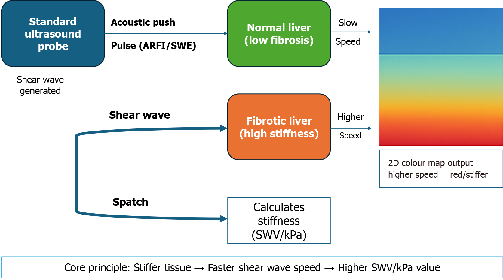
Figure 7 The principle of shear wave elastography for fibrosis assessment.
This schematic illustrates how shear wave elastography and acoustic radiation force impulse measure liver stiffness [shear wave velocity (SWV), measured in m/second or converted to kPa]. Unlike transient elastography, the shear wave is generated by a focused, acoustic push pulse emitted from a conventional ultrasound probe. This pulse perturbs the tissue, creating a shear wave that travels laterally. In normal liver tissue (low fibrosis), the wave travels slower, yielding a low SWV value. In fibrotic liver tissue (high stiffness), the wave travels faster. The system measures this speed and presents the result as a quantitative value (SWV) and often as a 2D color map, enabling real-time visualization of the stiffness distribution. This technique provides a non-invasive assessment of the inflammatory and fibrotic components of metabolic dysfunction-associated steatotic liver disease. ARFI: Acoustic radiation force impulse; SWE: Shear wave elastography; SWV: Shear wave velocity.
- Citation: Elbeltagi RM, Saeed NK, Bediwy AS, Al-Beltagi M. Ultrasound hepatic elastography: A non-invasive indicator of insulin resistance in the pediatric population: A systematic review. World J Clin Pediatr 2026; 15(2): 115963
- URL: https://www.wjgnet.com/2219-2808/full/v15/i2/115963.htm
- DOI: https://dx.doi.org/10.5409/wjcp.v15.i2.115963