
















Copyright: ©Author(s) 2026.
World J Clin Pediatr. Jun 9, 2026; 15(2): 115963
Published online Jun 9, 2026. doi: 10.5409/wjcp.v15.i2.115963
Published online Jun 9, 2026. doi: 10.5409/wjcp.v15.i2.115963
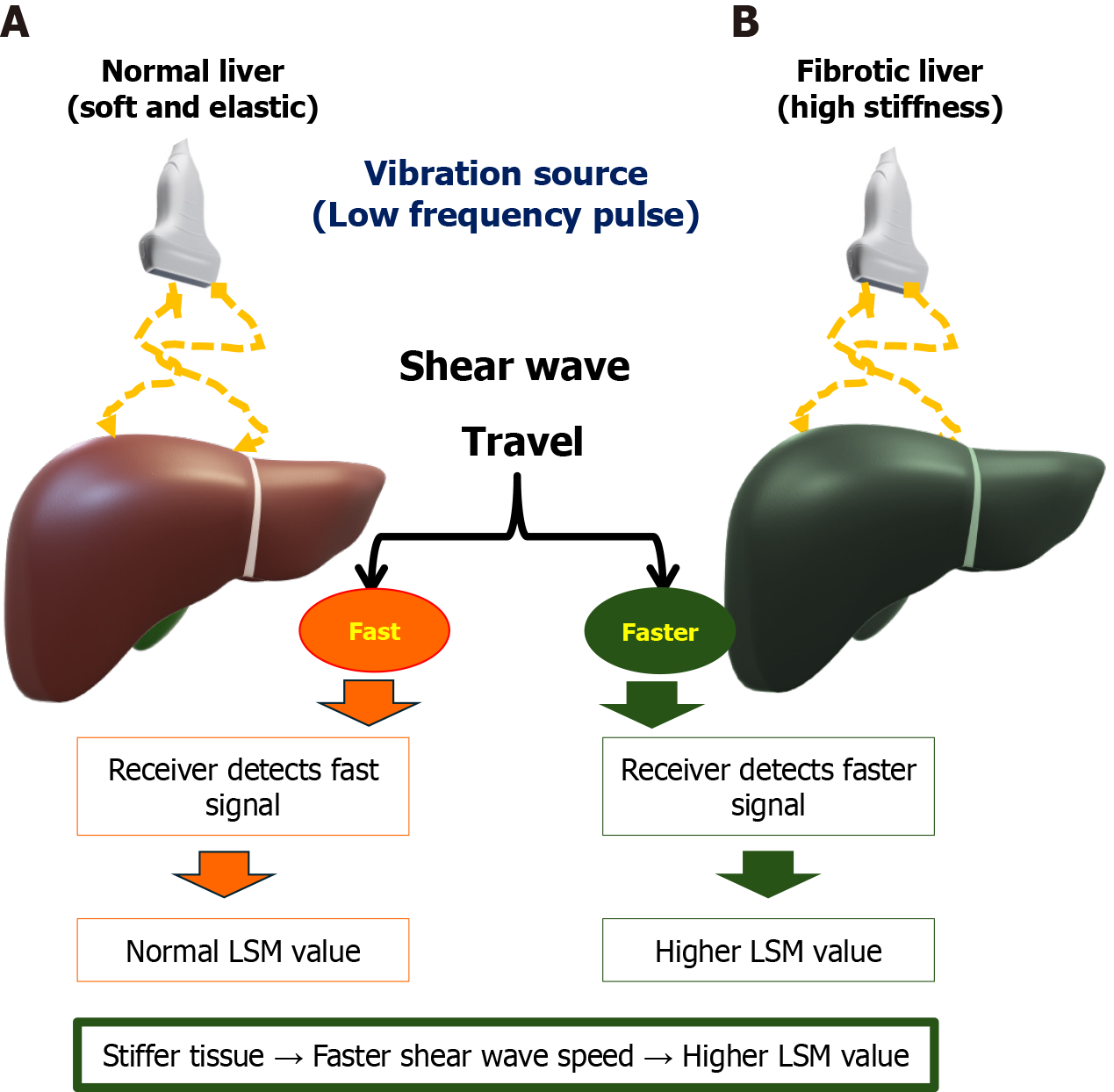
Figure 6 The principle of liver stiffness measurement for fibrosis assessment using transient elastography.
This schematic illustrates the mechanism by which liver stiffness measurement (LSM), typically measured in kPa, assesses liver fibrosis. LSM is derived from the speed of a low-frequency mechanical shear wave as it propagates through the liver. A: Depicts the shear wave traveling through a soft, normal (non-fibrotic) liver. The wave is minimally resisted and travels relatively slower; B: Depicts the shear wave traveling through a stiff, fibrotic liver (tissue hardened by collagen deposits). The rigid structure causes the shear wave to propagate faster. The core principle is that the stiffer the tissue, the faster the shear wave velocity. The LSM value is mathematically calculated from this velocity, providing a direct, non-invasive measure of liver stiffness, a validated surrogate for liver inflammation and fibrosis severity. LSM: Liver stiffness measurement.
- Citation: Elbeltagi RM, Saeed NK, Bediwy AS, Al-Beltagi M. Ultrasound hepatic elastography: A non-invasive indicator of insulin resistance in the pediatric population: A systematic review. World J Clin Pediatr 2026; 15(2): 115963
- URL: https://www.wjgnet.com/2219-2808/full/v15/i2/115963.htm
- DOI: https://dx.doi.org/10.5409/wjcp.v15.i2.115963