
















Copyright: ©Author(s) 2026.
World J Clin Pediatr. Jun 9, 2026; 15(2): 115963
Published online Jun 9, 2026. doi: 10.5409/wjcp.v15.i2.115963
Published online Jun 9, 2026. doi: 10.5409/wjcp.v15.i2.115963
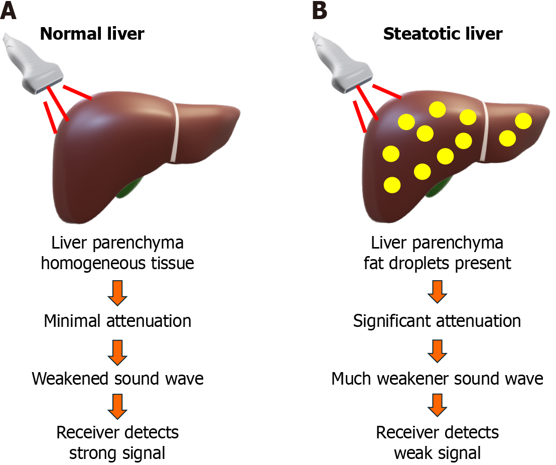
Figure 5 The principle of controlled attenuation parameter for steatosis assessment.
This schematic illustrates how the controlled attenuation parameter (CAP), measured in decibels per meter (dB/m), quantifies hepatic steatosis (liver fat). A: Sound waves passing through a healthy liver with homogeneous tissue, resulting in minimal signal energy loss (low attenuation) and a corresponding low CAP value; B: The effect of steatosis. Fat droplets (lipid vacuoles) are highly scattering and absorptive to the ultrasound signal. As the wave travels through the fatty liver tissue, it loses substantial energy, resulting in a significantly weakened signal upon returning to the receiver (high attenuation). This signal loss correlates directly with the degree of steatosis, yielding a high CAP value. CAP is therefore a direct, non-invasive biomarker for the physical presence of fat in the liver.
- Citation: Elbeltagi RM, Saeed NK, Bediwy AS, Al-Beltagi M. Ultrasound hepatic elastography: A non-invasive indicator of insulin resistance in the pediatric population: A systematic review. World J Clin Pediatr 2026; 15(2): 115963
- URL: https://www.wjgnet.com/2219-2808/full/v15/i2/115963.htm
- DOI: https://dx.doi.org/10.5409/wjcp.v15.i2.115963