
















Copyright: ©Author(s) 2026.
World J Clin Pediatr. Jun 9, 2026; 15(2): 114310
Published online Jun 9, 2026. doi: 10.5409/wjcp.v15.i2.114310
Published online Jun 9, 2026. doi: 10.5409/wjcp.v15.i2.114310
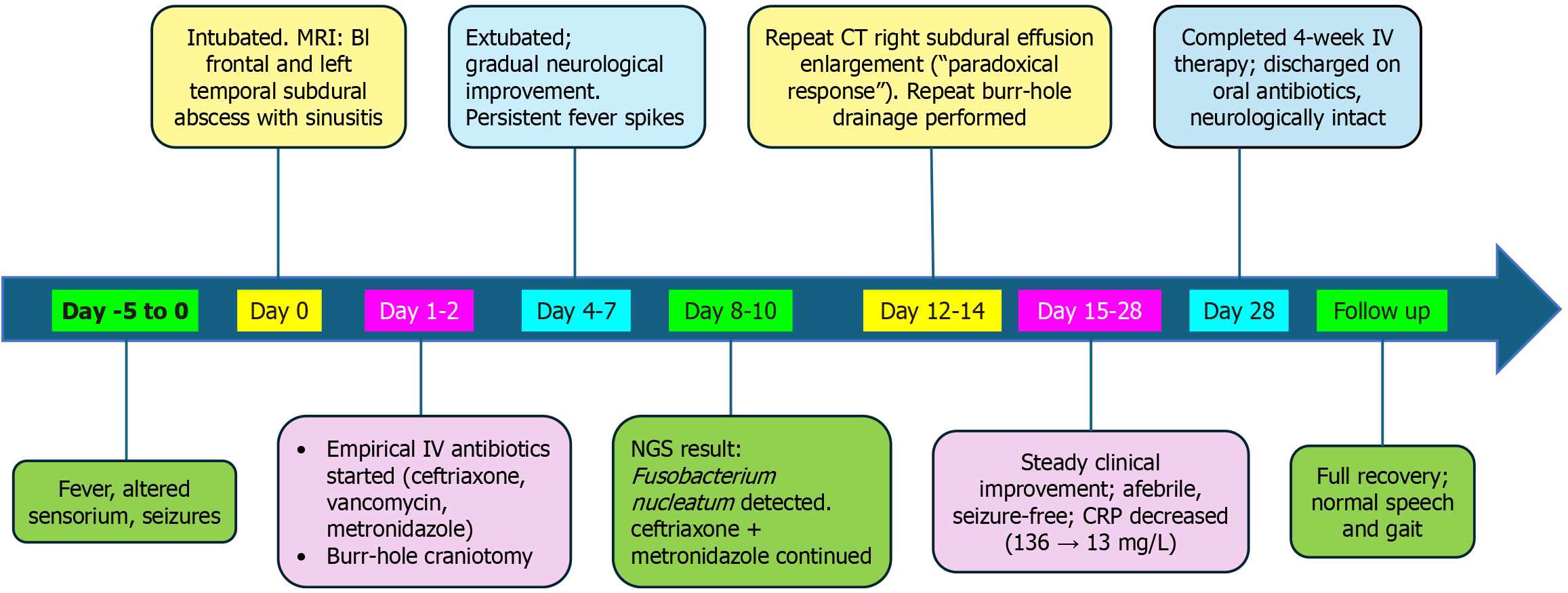
Figure 2 Clinical timeline of presentation, management, and recovery in a child with Fusobacterium nucleatum brain abscess secondary to sinusitis.
Day -5 to 0: Fever, headache, and altered sensorium led to magnetic resonance imaging showing bilateral frontal and left temporal subdural abscess with sinusitis. Day 0-2: Empirical ceftriaxone, vancomycin, and metronidazole were initiated; burr-hole drainage performed. Day 4-7: Gradual neurological improvement, persistent fever. Day 8-10: Next-generation sequencing confirmed Fusobacterium nucleatum; vancomycin stopped, ceftriaxone and metronidazole continued. Day 12-14: Repeat computed tomography showed paradoxical effusion enlargement; repeat drainage performed. Day 15-28: C-reactive protein decreased (136 mg/L→13 mg/L); afebrile, seizure-free. Follow-up: Completed 4-week IV antibiotics; discharged neurologically intact with normal speech and gait. MRI: Magnetic resonance imaging; CT: Computed tomography; IV: Intravenous; NGS: Next-generation sequencing; CRP: C-reactive protein; BI: Bilateral.
- Citation: Sankar J, Haribabu PPK, Singaravelu Suganya AGK. Fusobacterium brain abscess as a complication of sinusitis in an immunocompetent adolescent: A case report. World J Clin Pediatr 2026; 15(2): 114310
- URL: https://www.wjgnet.com/2219-2808/full/v15/i2/114310.htm
- DOI: https://dx.doi.org/10.5409/wjcp.v15.i2.114310