
















Copyright: ©Author(s) 2026.
World J Clin Pediatr. Jun 9, 2026; 15(2): 114310
Published online Jun 9, 2026. doi: 10.5409/wjcp.v15.i2.114310
Published online Jun 9, 2026. doi: 10.5409/wjcp.v15.i2.114310
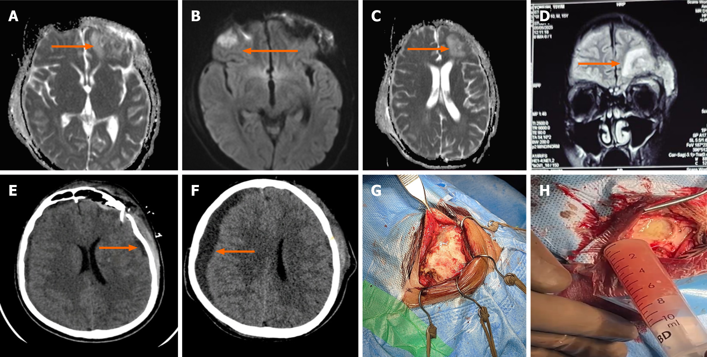
Figure 1 Neuroimaging and intraoperative findings in a 15-year-old male with Fusobacterium nucleatum brain abscess secondary to sinusitis.
Magnetic resonance imaging (MRI) brain demonstrating bilateral frontal subdural empyema and left frontal sinusitis. A: Axial T2-weighted image showing left frontal subdural collection with mass effect (arrow); B: Axial fluid-attenuated inversion recovery sequence revealing hyperintense collection and midline shift (arrow); C: Axial T1 post-contrast image showing ring enhancement of the abscess cavity (arrow); D: Coronal MRI depicting frontal sinusitis with contiguous spread to the left frontal lobe (arrow); E and F: Computed tomography (CT) brain showing postoperative findings. CT performed on day 4 revealing residual left frontotemporal effusion (arrow) (E). Repeat CT on day 10 demonstrating paradoxical enlargement of right-sided subdural effusion (arrow) following effective antibiotic therapy (F); G and H: Intraoperative images during burr-hole craniotomy showing exposure of the left frontal bone and aspiration of thick purulent material (approximately 100 mL). MRI: Magnetic resonance imaging; CT: Computed tomography.
- Citation: Sankar J, Haribabu PPK, Singaravelu Suganya AGK. Fusobacterium brain abscess as a complication of sinusitis in an immunocompetent adolescent: A case report. World J Clin Pediatr 2026; 15(2): 114310
- URL: https://www.wjgnet.com/2219-2808/full/v15/i2/114310.htm
- DOI: https://dx.doi.org/10.5409/wjcp.v15.i2.114310