
















Copyright: ©Author(s) 2026.
World J Clin Pediatr. Jun 9, 2026; 15(2): 114270
Published online Jun 9, 2026. doi: 10.5409/wjcp.v15.i2.114270
Published online Jun 9, 2026. doi: 10.5409/wjcp.v15.i2.114270
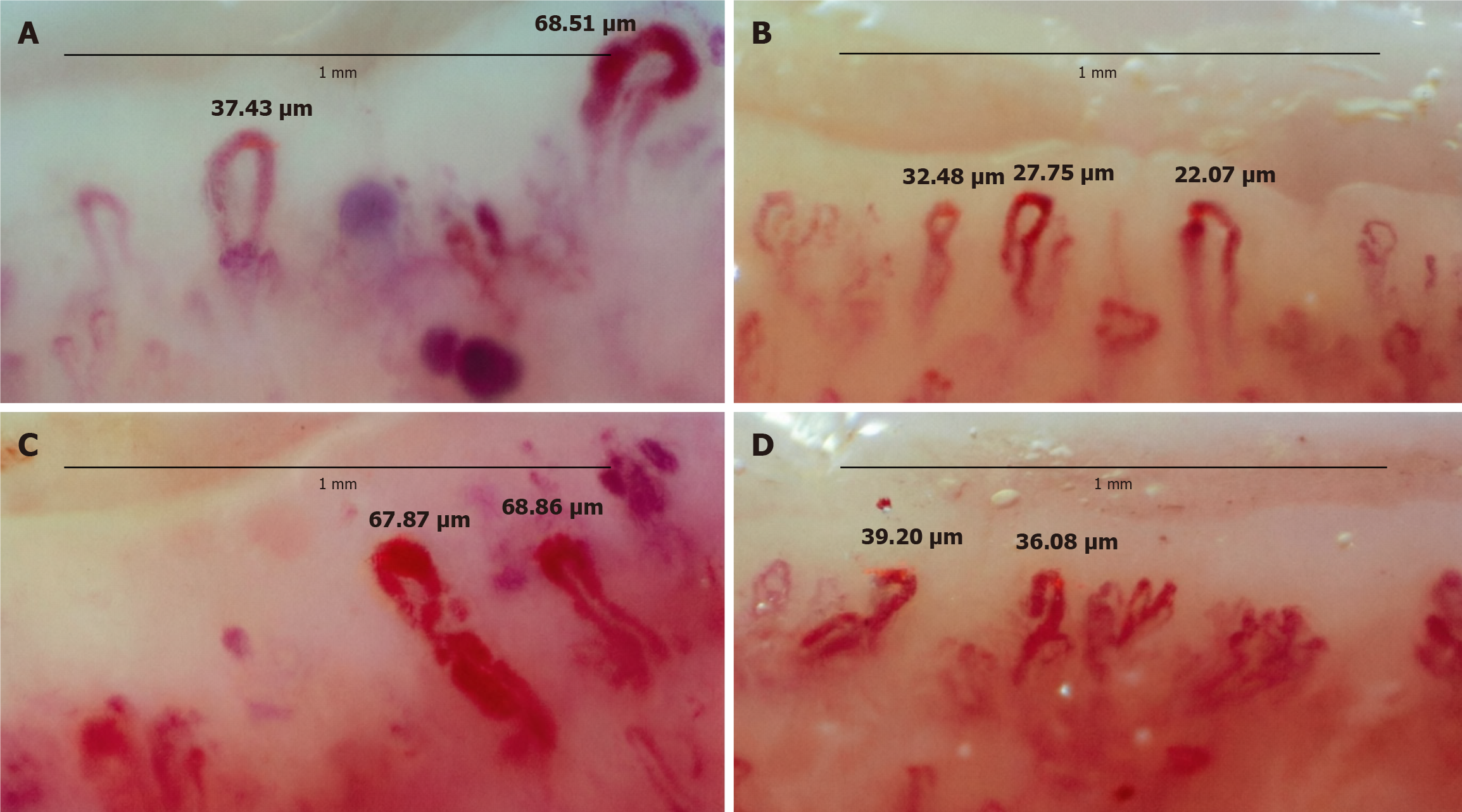
Figure 3 Dynamics of microvascular changes in a 15-year-old girl with juvenile dermatomyositis before and after therapy.
The black line at the top of each image represents 1 mm. Note the increase in capillary density from 2-3/mm to 4-5/mm, the disappearance of microhemorrhages and giant capillaries, and the emergence of a bushy capillary pattern, indicating neovascularization. A: Left hand, second finger before therapy; B: Left hand, second finger after four months of Janus kinase inhibitor therapy; C: Right hand, second finger before therapy; D: Right hand, second finger after 4 months of Janus kinase inhibitor therapy.
- Citation: Podzolkova V, Avrusin IS, Nikolaeva M, Afonina E, Davtian S, Nurseitova A, Kravtsova K, Malahova A, Yakovlev AA, Avrusin SL, Kalashnikova OV, Chasnyk VG, Kostik MM. Use of nailfold capillaroscopy for evaluation of disease activity in juvenile dermatomyositis: Results of a two-center retrospective study. World J Clin Pediatr 2026; 15(2): 114270
- URL: https://www.wjgnet.com/2219-2808/full/v15/i2/114270.htm
- DOI: https://dx.doi.org/10.5409/wjcp.v15.i2.114270