
















Copyright: ©Author(s) 2026.
World J Clin Pediatr. Jun 9, 2026; 15(2): 114189
Published online Jun 9, 2026. doi: 10.5409/wjcp.v15.i2.114189
Published online Jun 9, 2026. doi: 10.5409/wjcp.v15.i2.114189
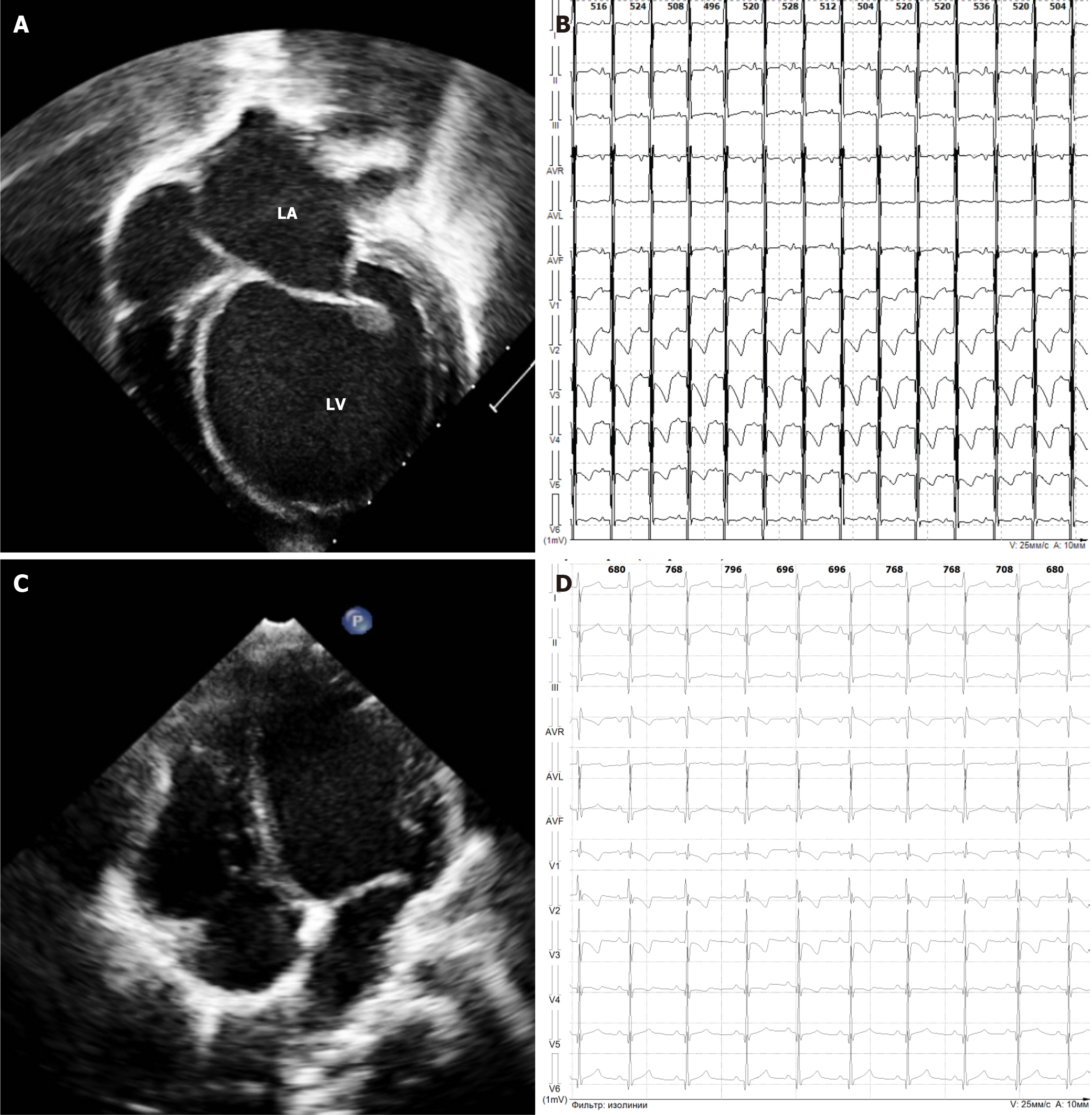
Figure 5 Echocardiography and Electrocardiography findings: A and B: At 3 months of age: Left ventricular dilation with spherical configuration and diffuse repolarization abnormalities, including T-wave inversion on electrocardiography (ECG); C and D: At 2 years of age: Normalization of left ventricular size (LVEDD 24.
5 mm; Z-score +1.96) and resolution of T-wave inversion with normalized repolarization patterns on ECG.
- Citation: Melnik OV, Kulichik OE, Zaytseva AK, Kofeynikova OA, Fetisova SG, Tarnovskaya SI, Fomicheva YV, Vasichkina ES, Zhorov BS, Kalinina OV, Kostareva AA. Role of TRPM4 ion channel in pediatric arrhythmic syndromes. World J Clin Pediatr 2026; 15(2): 114189
- URL: https://www.wjgnet.com/2219-2808/full/v15/i2/114189.htm
- DOI: https://dx.doi.org/10.5409/wjcp.v15.i2.114189