
















©Author(s) (or their employer(s)) 2026.
World J Clin Pediatr. Mar 9, 2026; 15(1): 110318
Published online Mar 9, 2026. doi: 10.5409/wjcp.v15.i1.110318
Published online Mar 9, 2026. doi: 10.5409/wjcp.v15.i1.110318
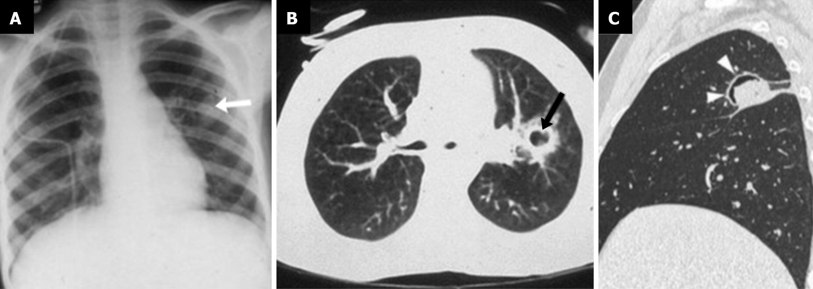
Figure 3 Aspergillus infection in a child[7].
A: Chest X-ray showcases a solitary rounded pulmonary lesion in a patient with leukemia (arrow); B: Further evaluation with computed tomography demonstrates cavitation of the lesion and a ground glass halo sign (arrow); C: Different immunocompromised patient with fungal infection showcasing the "air crescent sign" (arrowheads). Citation: Varotto A, Orsatti G, Crimì F, Cecchin D, Toffolutti T, Zucchetta P, Stramare R. Radiological Assessment of Paediatric Fungal Infections: A Pictorial Review With Focus on PET/MRI. In Vivo 2019; 33: 1727-1735. Copyright © 2019 The Author(s). Published by the International Institute of Anticancer Research (https://iv.iiarjournals.org/content/editorial-policies).
- Citation: Schmiliver B, Perera Molligoda Arachchige AS. Imaging of pediatric pulmonary infections: A pictorial review. World J Clin Pediatr 2026; 15(1): 110318
- URL: https://www.wjgnet.com/2219-2808/full/v15/i1/110318.htm
- DOI: https://dx.doi.org/10.5409/wjcp.v15.i1.110318