
















©Author(s) (or their employer(s)) 2026.
World J Clin Pediatr. Mar 9, 2026; 15(1): 110318
Published online Mar 9, 2026. doi: 10.5409/wjcp.v15.i1.110318
Published online Mar 9, 2026. doi: 10.5409/wjcp.v15.i1.110318
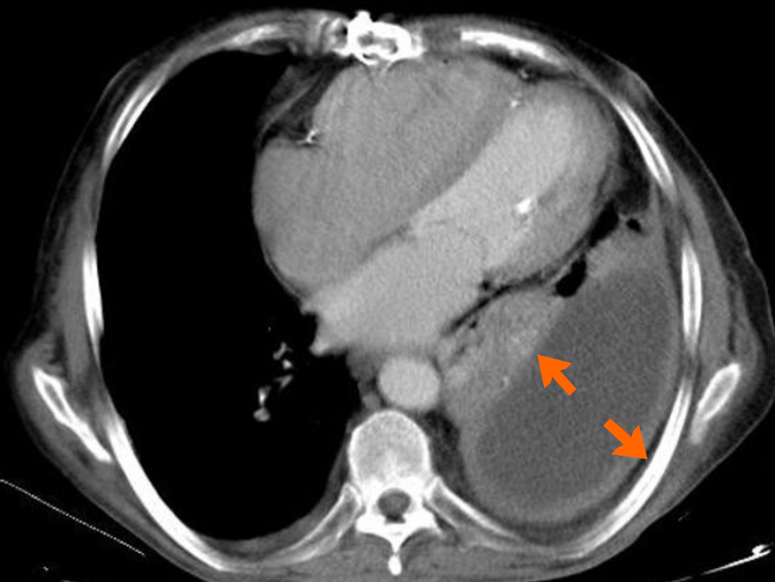
Figure 2 Axial contrast enhanced arterial phase computed tomography image showing the split pleura sign, seen with pleural empyemas refers to the thickening and increased contrast enhancement of the visceral and parietal pleura separated by empyema or an exudative effusion (arrows).
- Citation: Schmiliver B, Perera Molligoda Arachchige AS. Imaging of pediatric pulmonary infections: A pictorial review. World J Clin Pediatr 2026; 15(1): 110318
- URL: https://www.wjgnet.com/2219-2808/full/v15/i1/110318.htm
- DOI: https://dx.doi.org/10.5409/wjcp.v15.i1.110318