
















©The Author(s) 2025.
World J Clin Pediatr. Dec 9, 2025; 14(4): 108823
Published online Dec 9, 2025. doi: 10.5409/wjcp.v14.i4.108823
Published online Dec 9, 2025. doi: 10.5409/wjcp.v14.i4.108823
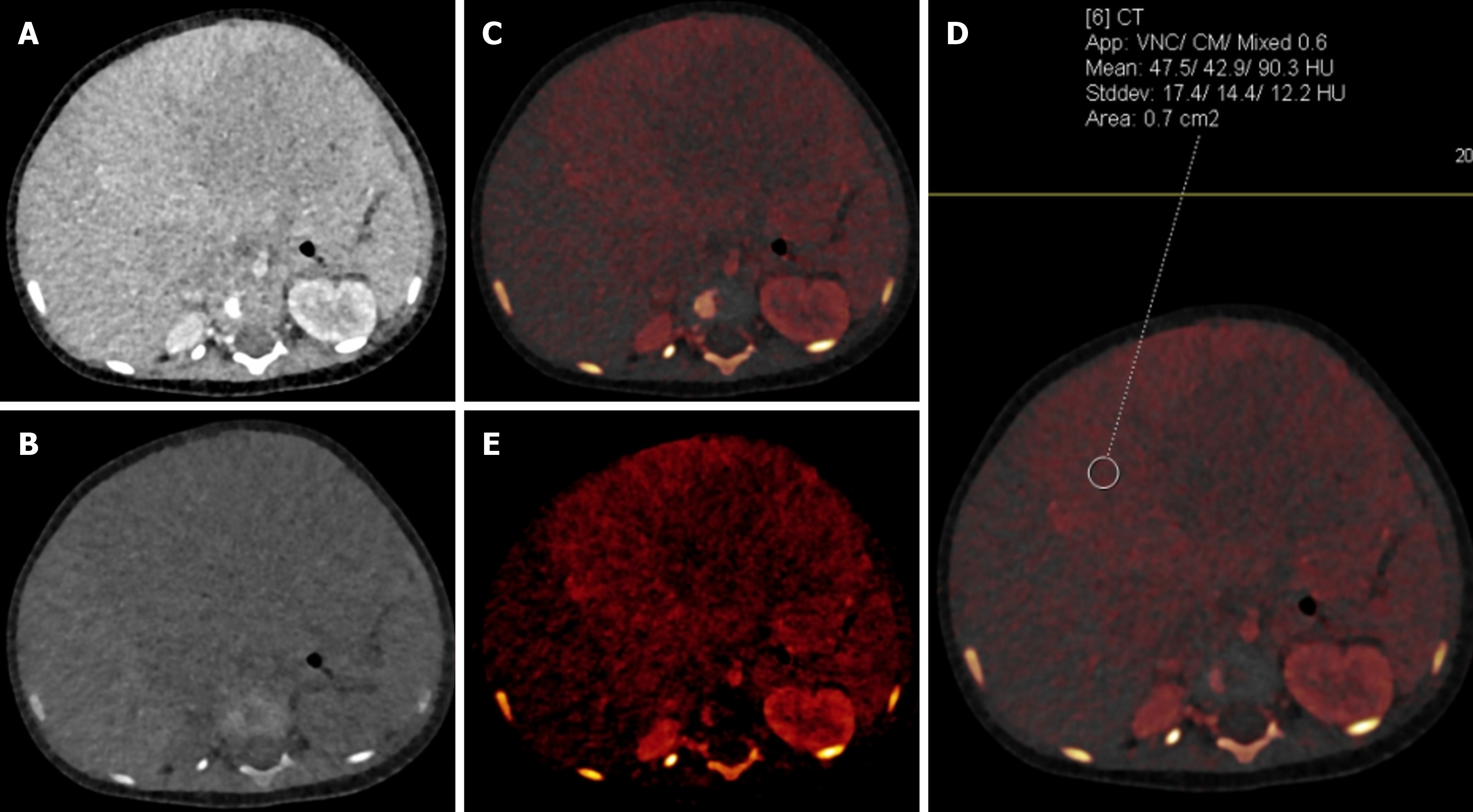
Figure 14 An 11-month-old male with hepatoblastoma.
A: Blended contrast-enhanced axial image showed a large isodense mass in the liver with poor demarcation of the mass with the adjacent liver due to minimal enhancement; B: Virtual non-contrast image showed no evidence of calcification/fat/hemorrhage within the lesion; C and D: Iodine overlay images showed iodine uptake in the periphery of the mass with attenuation on the virtual non-contrast image being 475 Hounsfield units and on the blended image being 903 Hounsfield units, suggesting significant enhancement that was not well appreciated on the blended image; E: Iodine-specific image showed the iodine content in the periphery of the mass to a better advantage.
- Citation: Saini S, Bhatia A, Bansal A, Saxena AK, Sodhi KS. Dual-energy computed tomography in children: Technique and clinical applications. World J Clin Pediatr 2025; 14(4): 108823
- URL: https://www.wjgnet.com/2219-2808/full/v14/i4/108823.htm
- DOI: https://dx.doi.org/10.5409/wjcp.v14.i4.108823