
















©The Author(s) 2025.
World J Clin Pediatr. Dec 9, 2025; 14(4): 108823
Published online Dec 9, 2025. doi: 10.5409/wjcp.v14.i4.108823
Published online Dec 9, 2025. doi: 10.5409/wjcp.v14.i4.108823
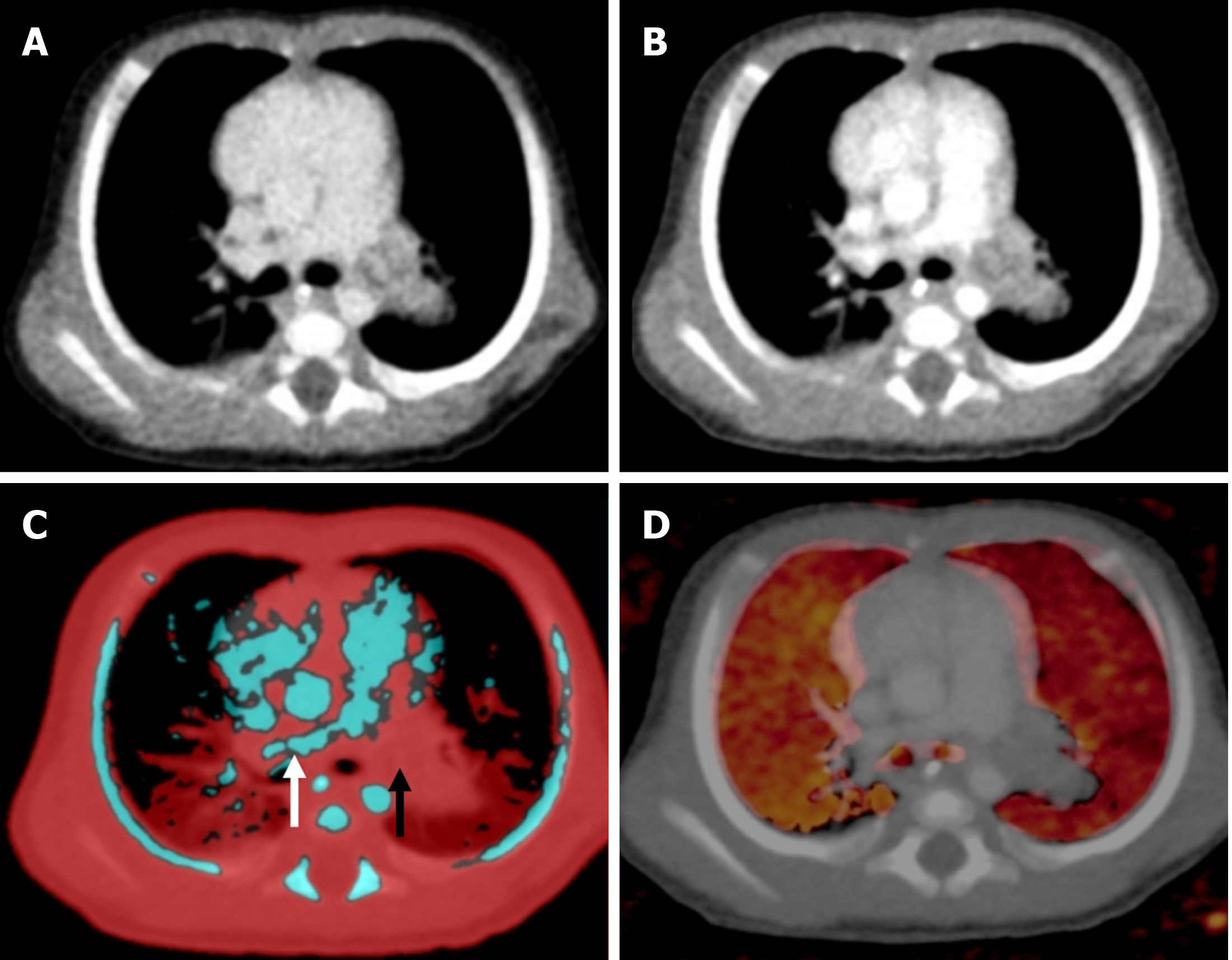
Figure 8 Axial sections of CT pulmonary angiography in a 2-month-old female with infective endocarditis.
A: Complete occlusion of the left pulmonary artery by a thrombus on blended contrast image; B: Complete occlusion of the left pulmonary artery by a thrombus on low kV (80 kV) image, showing increased contrast in the vasculature; C: Lung vessel image showed blue color-coded right pulmonary artery (white arrow) with thrombus being color-coded red in the left pulmonary artery (black arrow), suggesting diminished iodine content; D: Lung blood volume image showed diffusely reduced perfusion in the left lung as compared with the right lung.
- Citation: Saini S, Bhatia A, Bansal A, Saxena AK, Sodhi KS. Dual-energy computed tomography in children: Technique and clinical applications. World J Clin Pediatr 2025; 14(4): 108823
- URL: https://www.wjgnet.com/2219-2808/full/v14/i4/108823.htm
- DOI: https://dx.doi.org/10.5409/wjcp.v14.i4.108823